Category: Medical Conditions and Diseases

Detailed references on the conditions treated at Seamless Medical Centers, covering causes, symptoms, how each is diagnosed, and current treatment options.

  • Painless Rectal Bleeding: Bright Red Blood, No Pain

    Painless Rectal Bleeding: Bright Red Blood, No Pain

    What Painless Bleeding Usually Looks Like

    The thing that unsettles people most about painless rectal bleeding is the contradiction at the center of it. There is blood — sometimes a startling amount of it — and yet nothing hurts. The absence of pain does not feel like reassurance. It feels like a missing piece of information.

    Most people describing painless rectal bleeding are seeing one of the following:

    • Bright red blood on the toilet paper, with no discomfort at any point
    • Blood dripping into the bowl after a bowel movement, turning the water red, with no pain
    • A surprisingly large amount of blood in the toilet, again with no pain
    • Bright red blood on the paper but nothing at all in the stool itself
    • Blood appearing only after a bowel movement, never at other times

    There is a good physiological reason all of these can happen without pain, and understanding it will probably settle a lot of your anxiety. It is also the reason painless bleeding still deserves a proper evaluation rather than a shrug — the same anatomy that makes it painless is what makes it easy to dismiss.

    Does Painless Bleeding Ever Need Urgent Care?

    The absence of pain is not, by itself, an all-clear. Place yourself in one of the tiers below before reading further.

    Call 911 or go to an emergency room now if you have:

    • Heavy or continuous bleeding, even with no pain at all
    • Dizziness, lightheadedness, fainting, or weakness on standing
    • A racing heart, shortness of breath, or cold, clammy skin

    Arrange to be seen within 24 hours if you have:

    • Black, tarry, or maroon-colored stool
    • Painless bleeding while taking a blood thinner
    • Bleeding that is clearly increasing in volume each time

    Book an appointment if you have:

    • Painless bleeding still happening after one to two weeks, or recurring
    • Any rectal bleeding at 45 or older with no previous colonoscopy
    • Bleeding with a change in bowel habits, weight loss, fatigue, or narrower stools
    • A family history of colorectal cancer or inflammatory bowel disease

    Most people reading this fall into the third tier: bleeding, no pain, otherwise feeling fine. That is not an emergency — and it is also not something to keep living with indefinitely.

    Why Internal Hemorrhoids Bleed Without Hurting

    Inside the anal canal there is a boundary called the dentate line. It is not an arbitrary landmark. It marks where two entirely different types of tissue meet, and it is the key to this whole question.

    Below the dentate line, the tissue is derived from skin and is densely supplied with somatic nerve endings — the same kind of nerves that make a paper cut hurt. Anything that goes wrong down there is felt sharply and immediately. External hemorrhoids sit below the line. So do anal fissures. This is exactly why a thrombosed external hemorrhoid is excruciating and why a fissure produces that distinctive tearing, burning pain during a bowel movement.

    Above the dentate line, the tissue is rectal mucosa, and it is supplied by visceral nerves that register stretch and pressure but do not carry sharp pain. Internal hemorrhoids sit above the line. They can enlarge, become engorged, and have their fragile surface disrupted by passing stool — and you will not feel any of it. What you will see is blood.

    That is the whole explanation. Painless bright red rectal bleeding is not a strange or ominous combination of symptoms. It is the single most characteristic presentation of internal hemorrhoids, and it is precisely what you would predict from the anatomy. The bleeding is bright red because the source is only inches from the outside and the blood has not been digested. It is painless because the tissue it is coming from cannot generate that kind of pain.

    Understanding this also explains something people often find confusing: it is entirely possible to have significant internal hemorrhoids for years, bleeding intermittently, without ever having had a moment of hemorrhoid pain. The absence of pain has never been evidence that hemorrhoids are absent or mild.

    A Large Amount of Bright Red Blood in the Toilet With No Pain

    This is the version that sends people looking for answers at two in the morning, and it deserves a direct, honest answer.

    Blood disperses in toilet water dramatically. A relatively small volume — a few milliliters — can turn the entire bowl convincingly red and look, to anyone who has just produced it, like a medical emergency. This is a genuine and very common optical effect, and the amount of blood present is routinely far less than it appears.

    That said, volume is one of the things that does matter, and it is worth being clear about where the line sits. Bright red bleeding into the bowl after a bowel movement, in someone who feels otherwise entirely well, is most often hemorrhoidal, even when the bowl looks alarming. But bleeding that is heavy and continuous, bleeding that soaks through pads, bleeding accompanied by dizziness, lightheadedness, a racing heart, shortness of breath, weakness, or fainting, or bleeding that comes with severe abdominal pain — those are emergencies and they need immediate care, not an appointment next week. Significant ongoing blood loss can drop your blood pressure, and the symptoms above are how that presents.

    The middle ground is where most people actually are: a visible, sometimes substantial amount of blood, no pain, no dizziness, feeling otherwise fine. That situation is not an emergency, and it also is not something to keep watching indefinitely. It is a reason to be evaluated in the near term. Chronic hemorrhoidal bleeding, even at a low level, can quietly produce iron-deficiency anemia over months — which is one of the more practical arguments against simply living with it.

    Blood After Poop, but Nothing in the Stool

    The pattern of blood appearing after a bowel movement but never mixed into the stool itself is diagnostically useful, and it points in a reassuring direction.

    Blood that is mixed evenly through the stool joined it somewhere higher in the colon, before the stool was fully formed. That widens the field of possible causes considerably and is the finding most likely to prompt a colonoscopy. Blood that appears only afterward — on the paper, in the bowl, streaked on the outside of an otherwise normal stool — was picked up at the very end of the journey, in the anal canal or lower rectum. That is hemorrhoid and fissure territory.

    Combine that with the absence of pain and the picture narrows further. Painless, bright red, after-the-fact bleeding with a normal-looking stool is about as characteristic of internal hemorrhoids as a symptom pattern gets. If it hurt, a fissure would climb the list. If the blood were dark or mixed in, the source would likely be higher.

    None of which means the pattern is a diagnosis. It means it is a strong hypothesis that an evaluation can confirm in a few minutes. The reason to confirm it rather than assume it is straightforward: hemorrhoids are so common that having them tells you very little about whether you also have something else. An obvious explanation sitting in plain view is exactly the circumstance in which a second, less obvious cause gets missed.

    Painless Does Not Mean Harmless

    There is an assumption buried in a lot of searching around this symptom — that pain is the body’s alarm system, so no pain means no problem. It is worth taking that assumption apart, because in this particular part of the body it does not hold.

    The rectum and colon simply do not generate sharp pain the way skin does. Polyps do not hurt. Early colorectal cancers do not usually hurt. Neither, as established above, do internal hemorrhoids. The absence of pain is therefore not informative about severity — it is a fact about the anatomy of the region, and it applies equally to the benign causes and the ones you would rather rule out.

    This is the honest reason painless rectal bleeding warrants evaluation. Colorectal cancer can present as painless rectal bleeding, and it is worth knowing that incidence in adults under 50 has been rising, which is why routine screening now begins at 45 for adults at average risk rather than 50. Bleeding is not usually its only sign — a persistent change in bowel habits, stool that has become noticeably narrower, unexplained weight loss, fatigue out of proportion to your life, or a sense of never fully emptying tend to appear alongside it. The great majority of people with painless bright red bleeding have hemorrhoids, and they will be told so. But the only way to be told so is to be examined.

    If you are 45 or older, have never had a colonoscopy, and are now bleeding painlessly, that combination is the clearest possible reason to book the appointment. The same applies at any age if you have a family history of colorectal cancer or inflammatory bowel disease. Getting evaluated is not a sign you think something is wrong. It is how a benign explanation becomes a confirmed one.

    What Happens at an Evaluation

    The examination people dread turns out to be brief and undramatic. It begins with a conversation about the bleeding — how long, how often, how much, whether anything hurts, what your bowel habits have been doing, what you have already tried. That history does a surprising amount of the diagnostic work on its own.

    It is followed by an examination of the area and, commonly, an anoscopy: a short look inside the anal canal and lower rectum with a small lighted instrument. Internal hemorrhoids are visible directly this way, and they are graded by how far they prolapse, which is what determines the treatment options available to you. Depending on your age, your risk factors, and what is found, a colonoscopy may be recommended to examine the full length of the colon.

    The purpose is not just to find hemorrhoids. It is to confirm that the hemorrhoids are actually the source of your bleeding, and to make sure nothing is hiding behind them.

    When Painless Bleeding Keeps Coming Back

    For most people, bleeding from internal hemorrhoids improves with the unglamorous fundamentals: substantially more dietary fiber, more water, not straining, and not sitting on the toilet reading your phone for twenty minutes at a time. Those measures resolve a great deal of intermittent hemorrhoidal bleeding, and they are the right first step.

    When the bleeding keeps returning despite all of that — for months, for years, through every cream and every fiber supplement — the honest conclusion is that the underlying arterial supply feeding the hemorrhoidal tissue has not changed, and it is not going to change on its own. At that point the options are procedural. Rubber band ligation is frequently effective for lower-grade internal hemorrhoids. Surgical hemorrhoidectomy is definitive and has a well-earned reputation for a difficult recovery, because it involves cutting in tissue that — being below the dentate line — is exquisitely pain-sensitive.

    Hemorrhoid artery embolization sits between those two, and it follows directly from the anatomy discussed above. Internal hemorrhoids bleed because arteries are delivering excessive blood flow to fragile tissue. HAE addresses that supply directly: a thin catheter is guided from a small puncture in the wrist or groin to the arteries feeding the hemorrhoids, and their flow is selectively reduced, allowing the engorged tissue to shrink and the bleeding to settle. Critically, nothing is done to the anal canal itself. There is no incision in the pain-sensitive tissue below the dentate line, which is the reason recovery is so much lighter than after hemorrhoidectomy — most patients go home the same day and are back to normal activity within a few days.

    HAE is aimed at bleeding rather than at pain from external or thrombosed hemorrhoids, and it is not the right answer for everyone. But for the person whose defining symptom is exactly what this article is about — painless, recurrent, bright red bleeding from internal hemorrhoids that has outlasted every conservative measure — it is often the option nobody mentioned.

    At Seamless Medical Centers, Dr. Zagum Bhatti, Board-Certified Interventional Radiologist and Founder & Chief Medical Officer of Seamless Medical Centers, performs hemorrhoid artery embolization as a non-surgical treatment for hemorrhoid bleeding at the practice’s Port Arthur, Texas office. If you want the fuller picture of the symptom itself, our article on what blood when you wipe means and when to get it checked covers the full range of bleeding patterns. To weigh the procedures against each other, read our comparison of HAE and hemorrhoidectomy, and to understand the timeline afterward, our guide to recovery after hemorrhoid artery embolization walks through what to expect. For appropriate candidates, HAE may offer meaningful improvement in bleeding; individual results vary, and only an evaluation can determine whether you are a candidate. If you are also trying to work out why the hemorrhoids formed at all, understanding what causes hemorrhoids is a useful companion piece.

    Painless bleeding is one pattern among several. For the complete picture — why hemorrhoids bleed, what the color and pattern of the blood mean, and every treatment option from conservative care through embolization — see our full guide to hemorrhoid bleeding causes and treatment.

    Frequently Asked Questions About Painless Rectal Bleeding

    Why am I bleeding from my anus with no pain? Painless bright red rectal bleeding is most often caused by internal hemorrhoids. They sit above the dentate line, in tissue that has no sharp-pain nerve endings, so they can bleed noticeably without hurting. Painful bleeding is more suggestive of an anal fissure or an external hemorrhoid.

    I saw a large amount of bright red blood in the toilet but felt no pain. Is that an emergency? Blood disperses widely in toilet water, so the volume usually looks far greater than it is. If you feel otherwise well, it is not typically an emergency, though it does warrant evaluation soon. Seek immediate care if you have dizziness, lightheadedness, a racing heart, weakness, fainting, or continuous heavy bleeding.

    Can painless bleeding still be serious? Yes, which is why it should be evaluated. The colon and rectum do not produce sharp pain the way skin does, so the absence of pain says nothing about the underlying cause. Most painless bleeding is hemorrhoidal, but confirming that requires an examination rather than an assumption.

    Why is there blood when I wipe but not in my stool, and no pain? That combination points strongly to a source at the very end of the digestive tract — the anal canal or lower rectum — with internal hemorrhoids the most likely explanation. Blood mixed evenly through the stool suggests a source higher in the colon and generally prompts further investigation.

    What can be done if painless hemorrhoid bleeding keeps returning? Fiber, hydration, and avoiding straining resolve many cases. When bleeding persists despite that, options include office procedures such as rubber band ligation and catheter-based hemorrhoid artery embolization, which reduces the arterial supply feeding the hemorrhoids without any incision in the anal area. An evaluation determines which approach fits your situation.

    Talk to a Specialist About Painless Rectal Bleeding

    If you are bleeding without pain and it keeps coming back, an evaluation is what turns a likely explanation into a confirmed one. Contact Seamless Medical Centers to schedule a consultation with Dr. Bhatti and find out whether hemorrhoid artery embolization is an appropriate option for you.

    Phone: 409-213-9575 Address: 3300 Jimmy Johnson Blvd, Suite #130, Port Arthur, Texas 77642

    Medical Disclaimer

    Individual results may vary. This information is for educational purposes only and should not replace professional medical advice. Treatment decisions should be made in consultation with qualified healthcare providers.

    Published by Seamless Medical Centers. Clinical information reflects the expertise of Dr. Zagum Bhatti, MD, Board-Certified Interventional Radiologist and Founder & Chief Medical Officer of Seamless Medical Centers.

  • Hemorrhoid Artery Embolization: Non-Surgical Hemorrhoid Treatment

    Hemorrhoid Artery Embolization: Non-Surgical Hemorrhoid Treatment

    If you have been managing hemorrhoids for months or years and conservative treatments have stopped helping, you may be facing a recommendation for hemorrhoidectomy — surgical removal of hemorrhoids. Before committing to surgery, it is worth knowing that hemorrhoid artery embolization offers a non-surgical alternative that addresses hemorrhoid bleeding through a catheter-based procedure, with significantly less pain and a much faster recovery than traditional surgical approaches.

    Hemorrhoids are one of the most common conditions affecting adults, but the range of treatment options is not always clearly explained. Many patients progress from over-the-counter treatments to physician-administered office procedures to surgery without ever hearing about interventional radiology options that exist between those endpoints.

    At Seamless Medical Centers, Dr. Zagum Bhatti, Board-Certified Interventional Radiologist, performs HAE for patients across Southeast Texas and the Houston area. Both Houston-area HAE services and Port Arthur HAE services are available at our Port Arthur office.

    What Are Hemorrhoids and Why Do They Cause Bleeding?

    Hemorrhoids are enlarged or inflamed blood vessels in and around the rectum and anus. Internal hemorrhoids develop inside the rectum, above the dentate line, and are the type most commonly responsible for rectal bleeding — the bright red blood you may notice on toilet paper or in the toilet bowl. External hemorrhoids develop under the skin around the anus and more commonly cause pain, itching, and discomfort rather than bleeding.

    Internal hemorrhoids bleed because the thin rectal mucosa covering them is fragile and easily disrupted during bowel movements. As hemorrhoids enlarge, the small arteries supplying them deliver increased blood flow, making the vessels more prominent and bleeding more likely. This is the mechanism that HAE targets: by reducing the arterial blood supply to the hemorrhoidal tissue, the engorgement decreases and bleeding improves.

    Risk factors for symptomatic hemorrhoids include chronic straining during bowel movements, prolonged sitting (including on the toilet), low-fiber diets, pregnancy, obesity, and family history. Chronic diarrhea or constipation both contribute to hemorrhoid development and worsening.

    The HAE Procedure: How It Works

    Hemorrhoid artery embolization works by selectively blocking the small arteries supplying the internal hemorrhoidal tissue, reducing blood flow to the engorged vessels and allowing them to shrink. This is performed using a catheter — a thin, flexible tube — inserted through a small puncture in the wrist or groin and guided to the hemorrhoidal arteries using real-time X-ray imaging.

    The procedure uses micro-coils or embolic particles to block specific arterial branches supplying the hemorrhoids. This targeted approach reduces hemorrhoidal blood flow without disrupting blood supply to the surrounding rectal tissue. The procedure is performed under conscious sedation, not general anesthesia, and takes approximately 60 to 90 minutes. Most patients go home the same day.

    Unlike traditional hemorrhoid procedures — including hemorrhoidectomy, rubber band ligation, and sclerotherapy — HAE does not involve any direct manipulation of the hemorrhoidal tissue through the anus. This is the primary reason the post-procedure pain is dramatically lower than after surgical hemorrhoidectomy, which notoriously involves significant post-operative pain due to the sensitivity of the anal region.

    Who Is a Candidate for HAE?

    HAE is most appropriate for patients with grade II or III internal hemorrhoids who have persistent bleeding despite conservative management and office-based treatments. Patients who have tried and not found lasting relief from fiber supplementation, sitz baths, topical medications, and rubber band ligation are often the most appropriate candidates for HAE evaluation. Patients who are not surgical candidates due to medical comorbidities, or who simply prefer to avoid surgery, are also often good candidates.

    HAE is primarily indicated for hemorrhoid bleeding rather than pain. Patients whose primary complaint is pain from thrombosed external hemorrhoids or pain from prolapsed tissue may be better served by surgical approaches that directly address the external components. During the consultation, Dr. Bhatti evaluates whether HAE is the most appropriate option based on your hemorrhoid grade, symptom pattern, and prior treatment history.

    HAE Compared to Hemorrhoidectomy

    Hemorrhoidectomy — surgical excision of hemorrhoidal tissue — is the most definitive treatment for symptomatic hemorrhoids and produces excellent long-term results. However, it is also one of the most painful outpatient procedures performed, with recovery typically involving two to four weeks of significant anal pain, limited activity, and careful wound care. Most patients require prescription pain medication for the first week to two weeks. Read the detailed comparison of HAE vs hemorrhoidectomy for a full breakdown of how these approaches differ.

    HAE recovery is significantly lighter. Most patients experience mild discomfort and return to work within two to three days. The absence of anal incisions means there is no wound to care for and no risk of the anal stricture or fecal incontinence that can occasionally complicate surgical hemorrhoidectomy. For patients in Southeast Texas and the Houston area who cannot accommodate a multi-week recovery, HAE’s recovery profile represents a meaningful practical advantage.

    For patients who want to understand whether HAE is right for their hemorrhoid situation, contact Seamless Medical Centers to schedule a consultation. Visit our services overview for information on the full range of procedures.

    What Recovery After HAE Looks Like

    Because hemorrhoid artery embolization is performed through a small catheter rather than an incision in a sensitive area, recovery is generally far easier than most people expect from a hemorrhoid procedure. Most patients go home the same day and return to light activity within a day or two, often back to desk work within a few days. There is no surgical wound in the anal area to care for, which is one of the main reasons many patients describe the recovery as relatively comfortable compared with traditional hemorrhoid surgery. It is normal to have mild pelvic or lower-abdominal achiness for a short time, and your care team will explain what to expect and how to manage it. Because the embolization works by gradually reducing blood flow to the hemorrhoidal vessels, improvement in bleeding and other symptoms tends to develop over the following weeks rather than overnight, and individual results vary. Many people are surprised that a problem which has caused years of bleeding can be addressed without the prolonged, painful recovery they associate with hemorrhoid surgery, and that shorter downtime is often what makes treatment feasible for those who have put it off because they could not afford weeks away from work. If you want a fuller picture, our detailed guide to what to expect during HAE recovery walks through the timeline, and you should always follow the specific aftercare instructions your physician provides. Knowing in advance that the recovery is typically mild, and far removed from the difficult healing associated with cutting procedures, can itself make the decision to move forward feel less daunting.

    What to Expect at Your HAE Consultation

    Deciding whether HAE is right for you begins with an evaluation rather than an assumption. At your consultation, the physician reviews your symptoms and history, examines the area, and determines the type and severity of your hemorrhoids, since internal hemorrhoids that bleed are generally the most suitable for embolization. This assessment also helps rule out other causes of rectal bleeding, an important step before treating, because not all rectal bleeding comes from hemorrhoids. For appropriate candidates, the physician explains how the procedure would work in your specific case, what improvement is realistic, and how it compares with the other options you may be weighing. You will have the chance to ask about recovery, risks, and what the day of the procedure involves. It helps to come prepared: noting how long your symptoms have lasted, how often you bleed, what home measures you have already tried, and how the problem is affecting your daily life gives the physician a clearer starting point and makes the visit more productive. The goal is a clear, individualized recommendation. HAE suits some patients and not others, and an honest evaluation is what determines which group you are in, so if you are dealing with persistent or recurrent bleeding, this conversation is the practical first step toward relief that lasts. Many people delay out of embarrassment, but specialists treat hemorrhoids routinely and the conversation is straightforward and confidential, and choosing to be evaluated does not commit you to a procedure – plenty of people leave reassured that conservative measures are enough for now, with a clear plan for what to watch for.

    Schedule Your Consultation

    To learn more about your options, contact Seamless Medical Centers to schedule a consultation with Dr. Bhatti. Phone: 409-213-9575. Address: 3300 Jimmy Johnson Blvd, Suite #130, Port Arthur, Texas 77642.

    Medical Disclaimer

    Individual results may vary. This information is for educational purposes only and should not replace professional medical advice. Treatment decisions should be made in consultation with qualified healthcare providers.

    Published by Seamless Medical Centers | Clinical information reflects the expertise of Dr. Zagum Bhatti, MD, Board-Certified Interventional Radiologist, Founder of Seamless Medical Centers.

  • PAE Safety Profile: Understanding the Risks and Side Effects of Prostate Artery Embolization

    PAE Safety Profile: Understanding the Risks and Side Effects of Prostate Artery Embolization

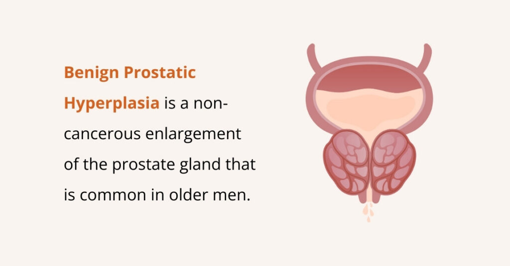
    When considering any medical procedure, understanding the safety profile — what side effects are common, what complications are possible, and how those compare to the alternatives — is essential for making an informed decision. Prostate artery embolization has an established safety record developed through years of clinical use, and its risk profile is substantially different from surgical alternatives for benign prostatic hyperplasia (BPH).

    At Seamless Medical Centers, Dr. Zagum Bhatti, Board-Certified Interventional Radiologist, performs PAE for patients with symptomatic BPH. Both Houston-area patients and Port Arthur patients are served at the Port Arthur office. Understanding the full safety picture is part of the consultation process.

    Common Temporary Side Effects

    PAE is associated with a well-characterized set of post-procedural effects that are expected, manageable, and temporary. Post-embolization syndrome — a cluster of symptoms including mild fever, pelvic discomfort or achiness, and general fatigue — is the most commonly reported set of effects in the days following PAE. These symptoms reflect the body’s normal inflammatory response to the embolization and typically resolve within three to five days.

    Urinary symptoms may temporarily worsen in the first one to two weeks after PAE before they improve. Urgency, frequency, and mild burning with urination are common in this early period. Patients who are not prepared for this transient worsening sometimes become concerned, but it is a recognized part of the early response to prostate embolization and does not indicate a problem. By the three to four week mark, most patients are experiencing improvement from their baseline.

    Mild discomfort at the catheter access site — typically the wrist or groin — is common and resolves within a few days. Bruising at the puncture site is also expected and normal.

    Serious Complications: Rare But Important to Understand

    Non-target embolization — the inadvertent delivery of embolic particles to arteries beyond the prostate — is the most significant potential complication of PAE. The prostate shares its blood supply with arteries supplying the rectum, bladder, and other pelvic structures. Careful technique using high-resolution imaging guidance and microspheres of the appropriate size minimizes this risk, but it cannot be eliminated entirely. In experienced hands, significant non-target embolization is uncommon.

    Urinary retention — inability to urinate — can occur in a small number of patients in the early post-procedural period as swelling in the prostate reaches its peak. This typically resolves within a few days with catheter placement. Urinary tract infection is also an occasional complication that is managed with antibiotics. Both are recognized, manageable events rather than unexpected complications.

    PAE’s Safety Advantages Over Surgical Alternatives

    Understanding PAE’s safety profile is most meaningful when compared to the alternatives. TURP — transurethral resection of the prostate — is the surgical standard and has well-documented side effects including retrograde ejaculation (in the majority of patients), sexual dysfunction, the risks of general or spinal anesthesia, and a recovery period measured in weeks. Open prostatectomy for very large prostates carries additional risks.

    PAE does not require general anesthesia — it is performed under conscious sedation. Sexual function, including ejaculatory function, is generally preserved after PAE, which is a significant advantage for sexually active men. Recovery is measured in days rather than weeks for most patients. For a detailed comparison of PAE and TURP across effectiveness, safety, and recovery, review the PAE vs TURP comparison.

    Who Is a Good Candidate From a Safety Perspective

    PAE is generally well-tolerated by patients who would otherwise face higher surgical risks due to medical comorbidities — including diabetes, cardiovascular disease, and chronic kidney disease — that make general anesthesia or surgical procedures higher-risk. The avoidance of general anesthesia and the minimally invasive nature of the catheter-based approach expand the candidate pool compared to surgical alternatives.

    Patients on blood thinners require coordination with their prescribing physician to temporarily adjust anticoagulation before the procedure. Patients with significant peripheral arterial disease affecting the iliac or femoral arteries may present technical challenges for access. These factors are evaluated during the pre-procedure consultation.

    If you have been diagnosed with BPH and are evaluating treatment options, understanding the full safety and effectiveness profile of PAE alongside other options helps you make the most informed decision. Review the PAE overview for a comprehensive introduction to the procedure. Contact Seamless Medical Centers to schedule a consultation. Visit our services page for information on all available treatments.

    Discussing PAE Safety at Your Consultation

    Your specific safety profile for PAE depends on factors that are evaluated individually during the pre-procedure consultation — including your prostate size, prostate artery anatomy, history of pelvic surgery or radiation, current medications, and underlying medical conditions. Larger prostates (above 80 grams) are technically feasible for PAE in experienced hands. A history of pelvic radiation may affect the arterial anatomy and procedural approach. Patients taking anticoagulants require specific preparation. None of these factors automatically preclude PAE, but each is part of the individualized assessment Dr. Bhatti performs before any procedure recommendation is made.

    Long-Term Safety Considerations

    Long-term follow-up data on PAE indicates a durable safety profile. The embolization is confined to the prostatic arteries, and surrounding structures maintain their blood supply through their own vascular networks. Sexual function is generally preserved because the pudendal arteries supplying the penile structures are not the target of the procedure. Ejaculatory function, which TURP frequently disrupts through retrograde ejaculation, is preserved in most PAE patients — one of the most meaningful safety and quality-of-life advantages for men who are sexually active.

    How the Risks of PAE Are Minimized

    Much of what makes PAE a well-tolerated procedure comes down to technique and planning. Before the procedure, imaging is used to map the arteries supplying the prostate, so the interventional radiologist understands each patient’s specific anatomy before any embolic material is delivered. During the procedure, high-resolution imaging guidance allows the catheter to be positioned precisely within the prostatic arteries, and embolic microspheres of a carefully chosen size are used to target the gland while sparing neighboring structures as much as possible.

    Patient selection is part of safety as well. The pre-procedure evaluation considers your prostate size, arterial anatomy, current medications, and overall health to determine whether PAE is a reasonable option for you and to anticipate any factors that warrant extra care. This individualized assessment is one reason an honest consultation matters: it allows the procedure to be tailored to your situation rather than applied as a one-size-fits-all approach.

    What to Watch For During Recovery

    Knowing what is normal after PAE makes the recovery far less stressful. Mild pelvic discomfort, low-grade fever, fatigue, and a temporary worsening of urinary symptoms are expected in the first days to weeks and generally settle on their own as the prostate responds. Over-the-counter measures and the guidance your care team provides are usually enough to manage this early period comfortably.

    It is equally important to know which symptoms warrant a call to your provider rather than watchful waiting. Difficulty urinating that does not improve, signs of infection such as a persistent or rising fever, significant bleeding, or pain that is worsening rather than easing should prompt you to reach out. As with any medical concern, symptoms that feel severe or alarming—or a complete inability to urinate—warrant prompt medical attention. Your care team will give you specific instructions on what to expect and when to call so you are never left guessing.

    Imaging, Contrast, and Radiation: Other Safety Considerations

    Because PAE is guided by X-ray imaging and uses iodinated contrast dye to visualize the arteries, two additional safety topics sometimes come up. The contrast material is processed by the kidneys, so your kidney function is reviewed beforehand, and steps are taken to protect the kidneys in men who have reduced function. If you have had a reaction to contrast in the past, that is important to share during your consultation so appropriate precautions can be taken.

    The procedure also involves a controlled dose of radiation from the imaging used to guide the catheter. In experienced hands, exposure is kept as low as reasonably achievable while still allowing the precise targeting that makes the procedure both safe and effective. For most men these considerations are routine and easily managed, but they are part of the complete safety picture worth discussing so you can make a fully informed decision.

    Your Next Step

    For men in Port Arthur, Beaumont, Nederland, Orange, and across the Golden Triangle and greater Houston area, the consultation at Seamless Medical Centers covers the full safety picture as it applies to your specific medical history and anatomy — including candidacy evaluation based on prostate size, symptom severity, and arterial anatomy. PAE is not appropriate for every patient with BPH, and part of the value of the consultation is an honest assessment of whether the procedure makes sense for your situation. Understanding what to expect, what is normal, and what to watch for after the procedure is central to how Dr. Bhatti approaches every patient interaction.

    If you have been diagnosed with BPH and are evaluating treatment options, understanding the full safety and effectiveness profile of PAE alongside other options helps you make the most informed decision. Review the PAE overview for a comprehensive introduction to the procedure. Learn more about PAE in Houston and PAE in Port Arthur. Contact Seamless Medical Centers to schedule a consultation with Dr. Bhatti.

    Phone: 409-213-9575

    Address: 3300 Jimmy Johnson Blvd, Suite #130, Port Arthur, Texas 77642

    Medical Disclaimer: Individual results may vary. This information is for educational purposes only and should not replace professional medical advice. Treatment decisions should be made in consultation with qualified healthcare providers.

    Medical Disclaimer

    Individual results may vary. This information is for educational purposes only and should not replace professional medical advice. Treatment decisions should be made in consultation with qualified healthcare providers.

    Published by Seamless Medical Centers | Clinical information reflects the expertise of Dr. Zagum Bhatti, MD, Board-Certified Interventional Radiologist, Founder of Seamless Medical Centers.
  • Hemorrhoid Bleeding Keeps Returning? When It’s Time to See a Specialist in Houston

    Hemorrhoid Bleeding Keeps Returning? When It’s Time to See a Specialist in Houston

    You’ve made the dietary changes. You’re drinking more water, taking fiber supplements, using over-the-counter creams. And for a while, it helps. But then, a few weeks or months later, the bleeding returns. Maybe it’s just a streak of bright red on the tissue, maybe it’s enough to color the bowl. Either way, you’re wondering: is this just something I’ll deal with forever, or is there a point where I should get this checked by a specialist?

    At Seamless Medical Centers, Board-Certified Interventional Radiologist Dr. Zagum Bhatti works with Houston-area patients—from Katy and Sugar Land to The Woodlands and League City—who’ve reached exactly this point. Recurring hemorrhoid bleeding doesn’t mean you’re out of options. It often means the condition has progressed to where conservative approaches alone aren’t enough, and more targeted treatment could help.

    Persistent or worsening rectal bleeding should be evaluated by a qualified healthcare provider—not just managed indefinitely with home remedies.

    Why Do Hemorrhoids Keep Bleeding?

    Hemorrhoids bleed because the swollen veins in the anal or rectal area become irritated during bowel movements. When stool passes over these inflamed tissues, the thin walls of the hemorrhoid can break open, causing bright red blood that appears on the tissue, in the bowl, or on the stool itself. For many people, this bleeding is intermittent—it stops on its own after a day or two, then returns weeks or months later.

    The cycle repeats because the underlying cause—the swollen hemorrhoid tissue itself—hasn’t been addressed. Dietary changes and creams can reduce irritation and make bowel movements easier, which may lessen bleeding temporarily. But if the hemorrhoid tissue remains enlarged and inflamed, it’s only a matter of time before symptoms return.

    Understanding what your rectal bleeding symptoms may indicate can help you recognize when you’ve moved beyond what home management can control.

    How Much Hemorrhoid Bleeding Is Normal?

    This is one of the most common questions patients ask, and the answer is nuanced. A small amount of bright red blood on the tissue after a bowel movement—especially if you’ve been constipated or straining—can occur with hemorrhoids and may not require immediate intervention. Many people experience this occasionally without needing treatment.

    However, several patterns suggest it’s time to seek evaluation. If the bleeding is frequent (happening with most bowel movements or several times a week), if the amount is increasing over time, if you’re seeing blood in the toilet bowl or significant staining on tissue, if the bleeding is accompanied by pain, swelling, or a feeling that something is protruding from the anus, or if you’re experiencing fatigue or lightheadedness that could indicate anemia from chronic blood loss, these are signals that warrant specialist assessment.

    It’s also important to note that while hemorrhoids are a very common cause of bright red rectal bleeding, they aren’t the only cause. Any persistent rectal bleeding should be evaluated to rule out other conditions.

    When Home Remedies Stop Being Enough

    Many Houston residents—whether you’re in Pearland, Missouri City, Cypress, or Spring—start managing hemorrhoid bleeding with conservative measures: increasing fiber, staying hydrated, using stool softeners, applying topical treatments. And for early-stage hemorrhoids, these approaches can be effective.

    But when bleeding returns despite these efforts, or when it becomes more frequent or heavier, that’s a signal the hemorrhoids have progressed to a point where they need more than dietary adjustments. You shouldn’t have to organize your life around avoiding flare-ups or live with the anxiety of wondering when the next bleeding episode will occur.

    Minimally Invasive Treatment for Bleeding Hemorrhoids

    When conservative treatment isn’t providing lasting relief, the traditional next step has been surgical hemorrhoidectomy—removal of the hemorrhoid tissue. While effective, surgery involves general anesthesia, significant post-operative pain, and a recovery period of several weeks.

    Hemorrhoid artery embolization (HAE) offers a different approach. Performed through a small catheter inserted at the wrist, HAE reduces blood flow to the hemorrhoid tissue, causing it to shrink. The procedure is done under light sedation, requires no incisions to the anal area, and most patients go home the same day. Learn how hemorrhoid artery embolization works and who it’s designed for.

    For appropriate candidates, HAE may provide substantial reduction in bleeding and other hemorrhoid symptoms. Recovery is significantly shorter than surgical options—read what HAE recovery involves to understand what each phase looks like. You can also compare HAE and hemorrhoidectomy to see how the approaches differ in recovery and outcomes.

    Accessing HAE in Houston

    Seamless Medical Centers provides hemorrhoid artery embolization for Houston-area patients throughout the metro. Whether you’re coming from Friendswood, Humble, Kingwood, Pasadena, or The Woodlands, the practice offers direct access to your treating physician from evaluation through follow-up.

    Unlike large hospital systems where you may see different providers at each visit, Seamless operates as a dedicated interventional radiology practice—focused exclusively on minimally invasive procedures. That means shorter wait times, continuity of care, and a team that specializes in exactly this type of treatment.

    When Recurrence Means It Is Time to See a Specialist

    There is a difference between hemorrhoid bleeding that flares once and settles, and bleeding that keeps coming back no matter how diligently you manage it – and recognizing which one you are dealing with is the heart of this decision. If you have genuinely given conservative measures a fair run – more fiber and water, not straining, topical treatments, sitz baths – over several weeks to a couple of months and the bleeding still returns, that recurring pattern is itself the signal. It tells you the problem is not a passing irritation but an enlarged hemorrhoid that keeps being disrupted, and that the underlying tissue, not your effort, is the limiting factor. A few specific patterns are worth treating as a clear prompt to seek specialist evaluation: bleeding that recurs every few weeks, episodes that are becoming heavier or more frequent rather than fading, blood that now fills the bowl rather than streaking the paper, or symptoms that have you planning around bathroom access and bracing for the next flare. Persistent blood loss, even in modest amounts, can occasionally contribute to anemia over time, which is another reason not to manage recurrent bleeding indefinitely. Seeing a specialist at this stage is not an overreaction – it is the logical next step once home care has shown you its limits.

    What Specialist Evaluation Involves

    If you have decided recurrent bleeding warrants a specialist, it helps to know that the visit is generally straightforward and aimed squarely at giving you answers. The physician reviews your history – how long the bleeding has been recurring, how heavy it gets, what you have already tried – and examines the area to identify the source and gauge the type and grade of your hemorrhoids. An important part of this step is confirming that hemorrhoids are in fact the cause and ruling out other explanations for recurrent bleeding, which is something assumption alone cannot do. From there, the conversation turns to what fits your situation: whether continued conservative management is reasonable, or whether a procedure makes sense given how persistent your symptoms have been. For internal hemorrhoid bleeding that has not responded to home care, this is often where minimally invasive options enter the picture, and you can come prepared by reading the broader overview of hemorrhoid symptoms and when treatment is warranted. Coming in with a sense of your pattern – frequency, amount, and what makes it worse – makes the visit more productive and helps your specialist tailor the recommendation. The point is not to push you toward any one treatment but to replace the cycle of recurrence with a clear, individualized plan. For many people who have spent months managing recurrent bleeding on their own, that single visit is also the moment the problem stops feeling open-ended, because they finally know what is causing it and what can be done about it.

    Frequently Asked Questions About Hemorrhoid Bleeding

    Can hemorrhoid bleeding stop on its own?

    Hemorrhoid bleeding often stops temporarily, especially after dietary changes or reduced straining. However, if the underlying swollen tissue remains, bleeding typically returns. If you’re experiencing a pattern of recurring bleeding, evaluation by a specialist can help determine whether more definitive treatment would be beneficial.

    Is bright red blood always from hemorrhoids?

    Bright red rectal bleeding is very commonly caused by hemorrhoids, but it’s not the only possible cause. Anal fissures, polyps, and other conditions can also produce bright red blood. Any persistent rectal bleeding warrants evaluation to confirm the cause and rule out other conditions.

    Will hemorrhoid artery embolization stop the bleeding?

    For appropriate candidates, HAE may provide significant reduction in hemorrhoid bleeding by shrinking the swollen tissue that’s causing symptoms. Many patients experience substantial improvement, though individual results vary based on the severity of the condition.

    How long does it take for hemorrhoid bleeding to improve after HAE?

    Many patients notice improvement in bleeding within the first few weeks following HAE as the hemorrhoid tissue begins to shrink. The full effect typically develops over one to two months. Your specialist will monitor your progress during follow-up appointments.

    Is hemorrhoid artery embolization covered by insurance?

    Many insurance plans cover HAE when it’s determined to be medically necessary. The team at Seamless Medical Centers can help coordinate with your insurance provider to understand your coverage before you commit to treatment.

    You Don’t Have to Keep Living With Bleeding

    If hemorrhoid bleeding has become a recurring problem despite your best efforts at home management, schedule your consultation with Seamless Medical Centers to discuss your symptoms and explore treatment options.

    Phone: 409-213-9575

    Address: 3300 Jimmy Johnson Blvd, Suite #130, Port Arthur, Texas 77642

    Medical Disclaimer

    Individual results may vary. This information is for educational purposes only and should not replace professional medical advice. Treatment decisions should be made in consultation with qualified healthcare providers.

    Published by Seamless Medical Centers | Clinical information reflects the expertise of Dr. Zagum Bhatti, MD, Board-Certified Interventional Radiologist, Founder of Seamless Medical Centers.
     Seamless Medical Centers home page

  • Non Surgical Fibroid Treatment for Long-Term Relief from Heavy Menstrual Bleeding

    Non Surgical Fibroid Treatment for Long-Term Relief from Heavy Menstrual Bleeding

    Heavy menstrual bleeding can significantly disrupt daily life, leading to fatigue, discomfort, and challenges at work or during social activities. For many women in Houston, uterine fibroids are a primary cause of prolonged or excessively heavy periods. While hysterectomy has long been viewed as a definitive treatment, many women now prefer less invasive alternatives that preserve the uterus, minimize recovery time, and offer long-term symptom relief. 

    Non-surgical fibroid treatments, such as Uterine Fibroid Embolization (UFE) and MRI-guided focused ultrasound, have proven effective in reducing fibroid size and controlling menorrhagia. At Seamless Medical Centers, women can access advanced, evidence-based therapies tailored to their unique needs, providing lasting relief from heavy menstrual bleeding while avoiding major surgery and promoting faster recovery. 

    Understanding Uterine Fibroids and Their Impact

    Uterine fibroids are noncancerous growths that develop in or around the uterus. They vary in size, number, and location, which influences the severity of symptoms.

    Common Symptoms of Fibroids

    • Heavy menstrual bleeding
    • Prolonged menstruation lasting more than 7 days
    • Pelvic pressure or pain
    • Frequent urination
    • Fatigue due to blood loss

    Among these, heavy bleeding is one of the most disruptive symptoms, often requiring specialized heavy bleeding treatment Houston.

    Why Consider Fibroid Treatment Without Surgery Houston

    Surgical procedures such as hysterectomy or myomectomy may not be suitable for everyone. Many women prefer fibroid treatment without surgery in Houston for several reasons.

    Benefits of Non Surgical Approaches

    • Preservation of the uterus
    • Shorter recovery time
    • Lower risk of complications
    • Outpatient or minimally invasive procedures
    • Faster return to normal activities

    These benefits make non surgical uterine fibroid treatments an increasingly preferred choice for women seeking effective yet less invasive care.

    Types of Non Surgical Fibroid Treatments Available

    There are several non-surgical options available today for managing fibroids. The most appropriate treatment depends on individual symptoms, fibroid size, location, and overall health. 

    Options such as uterine fibroid embolization, ovarian vein embolization, medication therapy, and minimally invasive procedures can provide effective symptom relief while reducing recovery time and preserving fertility, allowing women to maintain their daily activities with minimal disruption.

    1. Uterine Fibroid Embolization Houston

    Uterine fibroid embolization Houston is a minimally invasive procedure performed by interventional radiologists. It works by blocking the blood supply to fibroids, causing them to shrink over time.

    How It Works

    • A small catheter is inserted into a blood vessel
    • Tiny particles are used to block blood flow to fibroids
    • Fibroids shrink gradually, reducing symptoms

    Key Advantages

    • No surgical incision
    • Short hospital stay or same-day discharge
    • Effective for multiple fibroids
    • Significant reduction in heavy bleeding

    This procedure is widely recognized as a reliable menorrhagia treatment Houston.

    2. Hormonal Therapy for Symptom Control

    Hormonal medications can help regulate the menstrual cycle and reduce heavy bleeding.

    Common Options

    • Birth control pills
    • Hormonal intrauterine devices (IUDs)
    • Gonadotropin-releasing hormone agonists

    Limitations

    • Temporary relief
    • Symptoms may return after discontinuation
    • Not suitable for long-term use in all patients

    Hormonal therapy is often used as part of a broader prolonged menstruation treatment plan.

    3. MRI Guided Focused Ultrasound

    This is a non-invasive outpatient procedure that uses high intensity ultrasound waves to destroy fibroid tissue.

    Benefits

    • No incisions
    • Minimal recovery time
    • Targeted treatment

    Considerations

    • Not suitable for all fibroid types
    • Availability may be limited

    4. Endometrial Ablation

    Endometrial ablation removes or destroys the lining of the uterus to reduce bleeding.

    Suitable For

    • Women with smaller fibroids
    • Those not planning future pregnancies

    Limitations

    • Does not remove fibroids entirely
    • May not be effective for large or multiple fibroids

    Comparison of Non Surgical Fibroid Treatments

    Treatment OptionInvasivenessRecovery TimeEffectiveness for Heavy BleedingSuitable for Large Fibroids
    Uterine Fibroid EmbolizationMinimally invasiveShort 1 to 2 weeksHighYes
    Hormonal TherapyNon invasiveNoneModerateNo
    MRI Guided UltrasoundNon invasiveVery shortModerate to HighLimited
    Endometrial AblationMinimally invasiveShortModerateNo

    Choosing the Right Heavy Bleeding Treatment Houston

    Selecting the right treatment depends on several factors, including the severity of symptoms, underlying causes, overall health, and personal preferences. A personalized approach ensures the best outcomes, whether you are considering uterine fibroid embolization (UFE) or other advanced therapies, providing effective relief while minimizing risks and supporting long-term reproductive and overall health.

    Factors to Consider

    • Severity of symptoms
    • Size and number of fibroids
    • Age and reproductive goals
    • Overall health condition
    • Previous treatments

    Consulting a specialist experienced in uterine fibroid treatments can help determine the most suitable option.

    Long Term Relief from Menorrhagia

    Menorrhagia or excessive menstrual bleeding requires targeted care. Effective menorrhagia treatment Houston focuses on both symptom relief and addressing the root cause.

    Benefits of Treating Fibroids Early

    • Prevents anemia
    • Improves quality of life
    • Reduces risk of complications
    • Minimizes need for invasive surgery

    Early intervention using non surgical fibroid treatment Houston can provide lasting relief without major procedures.

    What to Expect During Treatment

    Before the Procedure

    • Medical evaluation and imaging tests
    • Discussion of treatment options
    • Personalized care plan

    During the Procedure

    • Minimally invasive or non invasive techniques
    • Typically performed under local or mild sedation

    After the Procedure

    • Short recovery period
    • Gradual improvement in symptoms
    • Follow up appointments for monitoring

    Patients undergoing uterine fibroid embolization Houston often notice reduced bleeding within a few menstrual cycles.

    Lifestyle and Supportive Measures

    While medical treatment is essential, lifestyle adjustments can support recovery and symptom management.

    Helpful Practices

    • Maintaining a balanced diet rich in iron
    • Regular physical activity
    • Stress management techniques
    • Tracking menstrual cycles

    These measures complement prolonged menstruation treatment and enhance overall well being.

    When to Seek Medical Advice

    It is important to consult a healthcare provider if you experience:

    • Bleeding lasting longer than 7 days
    • Passing large blood clots
    • Severe fatigue or dizziness
    • Pelvic pain or pressure

    Timely evaluation can help identify fibroids and initiate appropriate fibroid treatment without surgery Houston.

    Conclusion

    Non surgical fibroid treatment Houston offers effective less invasive solutions for women dealing with heavy menstrual bleeding and fibroids. With options like uterine fibroid embolization Houston and other advanced therapies it is possible to achieve long term relief without undergoing major surgery. 

    Understanding your options and seeking timely medical advice can help improve quality of life and support better long term health outcomes. Contact us today to schedule a consultation and explore the best treatment options for you.

    Frequently Asked Questions

    Q1. What is the most effective non surgical fibroid treatment in Houston

    Uterine fibroid embolization Houston is considered one of the most effective options for reducing fibroid size and controlling heavy bleeding without surgery.

    Q2. Can fibroids be treated without removing the uterus

    yes, many uterine fibroid treatments focus on preserving the uterus. Options like embolization and hormonal therapy are designed to manage symptoms while maintaining reproductive organs.

    Q3. How long does it take to recover from non surgical fibroid treatment

    Recovery time varies by procedure. Most minimally invasive treatments require a few days to two weeks which is significantly shorter than surgical recovery.

    Q4. Is non surgical treatment suitable for all fibroid cases

    Not all cases are the same. The suitability of fibroid treatment without surgery Houston depends on fibroid size location and patient health.

    Q5. Will heavy bleeding stop completely after treatment

    Many patients experience a significant reduction in bleeding. In some cases symptoms may resolve completely especially after uterine fibroid embolization Houston.

  • Fibroids and Pregnancy: Everything You Should Know

    Fibroids and Pregnancy: Everything You Should Know

    For many expecting parents, the discovery of fibroids during a routine prenatal ultrasound comes as a surprise.

    Fibroids are incredibly common, and in many cases, people don’t even know they have them until pregnancy brings them to light.

    As more individuals choose to start families later in life, when fibroids naturally become more frequent, these findings are becoming part of the modern pregnancy experience.

    While most pregnancies involving fibroids progress smoothly, their presence can raise understandable questions.

    • Will they affect the baby’s growth?
    • Increase the risk of preterm birth?
    • Lead to pain or complications later in pregnancy?

    The answers depend on factors like size, number, and location, and with the right monitoring, many people navigate pregnancy with fibroids without major issues.

    In this guide, we’ll explain the impact of fibroids on pregnancy, outline potential risks, describe diagnostic approaches, and provide practical strategies for managing fibroids while pregnant.

    How Fibroids Affect Pregnancy

    how fibroids affect pregnancy

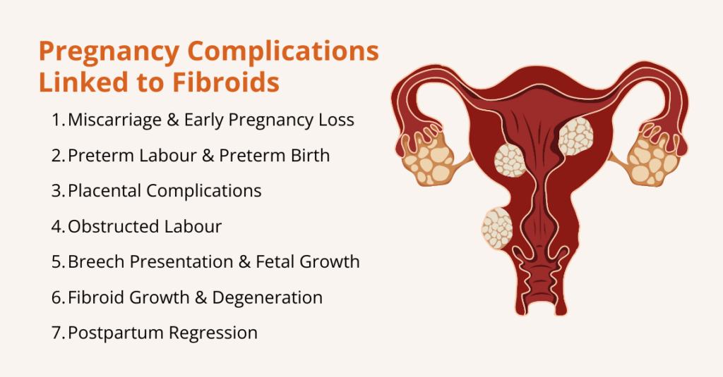
    The presence of uterine fibroids (even when they’ve been harmless before pregnancy) can influence pregnancy in several ways, from early pregnancy loss to complications late in pregnancy or at delivery. The risks vary based on fibroid size, number, and location.

    Here are some of the ways fibroids can influence pregnancy:

    1.  Miscarriage and Early Pregnancy Loss

    Fibroids, particularly submucosal lesions, are associated with an increased risk of miscarriage.

    A meta‑analysis pooling data from more than 237,000 participants found that women with fibroids had a higher risk of miscarriage than those without fibroids (relative risk RR ≈ 4.5, with miscarriage rates 13.42 % vs. 2.84 % in controls).

    The risk is highest when fibroids distort the uterine cavity, because the altered blood supply and mechanical interference impair implantation and early placental development.

    2. Preterm Labour and Preterm Birth

    Large or multiple fibroids can irritate the uterus and trigger contractions, leading to preterm labour.

    A study reported that preterm birth occurred in 12.85 % of pregnant patients with fibroids versus 9.43 % of controls; after adjusting for confounders, fibroids remained associated with preterm birth.

    Another study estimated that 10 %–30 % of pregnant women with fibroids develop obstetric complications such as preterm labour. Also, premature uterine contractions may be due to local inflammation, degenerating fibroids, or increased uterine irritability.

    3. Placental Issues

    Fibroids can interfere with placentation. If a fibroid is located near the placenta, it may lead to:

    • Placenta Previa: The placenta covers the cervix, obstructing the birth canal.
    • Placental Abruption: Premature separation of the placenta. The meta‑analysis found an elevated risk of placental abruption in women with fibroids.
    • Placenta Accreta Spectrum: Fibroids may increase the likelihood of abnormal placental attachment; however, the data remain limited.

    4. Need for Caesarean Delivery

    Large intramural or subserosal fibroids can physically obstruct the birth canal, preventing the fetal head from descending. Thus, fibroids raise the odds of cesarean delivery.

    For example, fibroids growing low in the uterus or near the cervix may block the birth canal, preventing the baby’s head from descending.

    Others may distort the uterus, making it harder for the baby to settle into a head-down position and leading to breech or transverse presentations.

    5. Breech Presentation and Fetal Growth Restriction

    Fibroids, especially those in the lower uterine segment, can distort the uterine cavity and limit space for the fetus. Research highlights that breech presentation or other fetal malposition is more common when fibroids are present, likely because fibroids can alter the shape of the uterine cavity.

    Similarly, in analyses comparing by fibroid size, larger fibroids conferred especially high risk of breech presentation and placenta previa.

    However, the overall effect on fetal growth remains uncertain because many pregnancies with fibroids result in normal birth weights.

    6. Fibroid Growth and Degeneration during Pregnancy

    Fibroids behave unpredictably during pregnancy.

    A study describes a woman in early pregnancy who arrived with severe abdominal pain, which imaging later confirmed was caused by a degenerating fibroid. This degeneration occurs when a fibroid outgrows its blood supply, leading to tissue ischemia and necrosis.

    7. Postpartum Regression

    After delivery, many fibroids shrink as hormonal levels decline and the uterus involutes.

    A case series found that fibroids decreased in size in 72% of women postpartum and that their volume could shrink by 50% within 6 months.

    Ongoing breastfeeding may further promote fibroid regression. Another observational study reported that 37 % of fibroids disappeared postpartum and that breastfeeding was associated with fibroid regression.

    What are the Risks and Complications of Fibroids during Pregnancy

    what are the risks and complications of fibroids during pregnancy

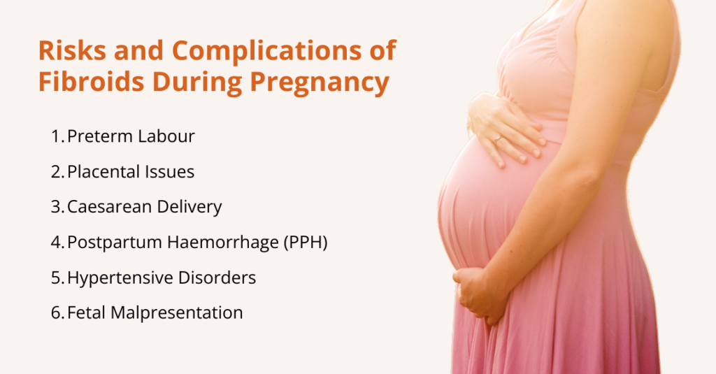
    While many women with fibroids have healthy pregnancies, these benign growths can sometimes create challenges as the uterus expands and hormonal changes intensify.

    Here are the key complications associated with fibroids during pregnancy:

    1. Premature Rupture of Membranes

    Fibroids can cause uterine irritability, increasing the frequency of contractions and elevating the risk of preterm labour.

    The high‑risk pregnancy resource notes that adverse outcomes such as preterm labour and premature rupture of membranes (PROM) are more common when fibroids exceed 10 cm.

    The meta‑analysis reported that fibroids were associated with both preterm birth and preterm PROM. Continuous antenatal care with ultrasound monitoring can help detect early cervical changes and manage preterm labour, sometimes using medications such as tocolytics to delay delivery.

    2. Heavy Bleeding Due to Placental Problem

    Placental problems can cause heavy bleeding and require early delivery or caesarean section. Women with placenta previa are more likely to deliver via caesarean section.

    Research shows that placenta previa may manifest as painless vaginal bleeding, often requiring bed rest or early delivery; placental abruption presents with painful bleeding and is an obstetric emergency.

    Importantly, fibroids located near the placenta or in the lower uterine segment increase these risks.

    3. Obstructed Labour

    Large fibroids can obstruct the cervix or lower uterine segment. When the fibroid blocks the birth canal, a caesarean delivery becomes necessary.

    Additionally, fibroids may cause dystocia (slow cervical dilation), prolonging labour and increasing the likelihood of operative delivery.

    The High‑Risk Pregnancy Information site states that a caesarean section is six times more likely in women with fibroids, highlighting the need for careful intrapartum planning.

    4. Postpartum Haemorrhage (PPH)

    PPH is excessive bleeding after delivery, often due to uterine atony (failure of the uterus to contract). Fibroids can impede uterine contraction, particularly if they distort the uterine muscle or occupy the uterine wall.

    The meta‑analysis found that postpartum haemorrhage occurred in 10.10 % of women with fibroids compared with 3.96 % in controls. Obstetric teams anticipate this risk and prepare for active management of the third stage of labour in women with known fibroids.

    5. Preeclampsia and Other Hypertensive Disorders

    Emerging evidence indicates a modestly increased risk of preeclampsia (new‑onset hypertension with end‑organ damage) in women with fibroids. The meta‑analysis reported an association between fibroids and preeclampsia.

    Although the absolute increase is small, pregnant women with fibroids should be monitored for blood pressure changes.

    6. Fetal Malpresentation and Growth Issues

    Fibroids can affect how a baby positions itself in the uterus, sometimes leading to malpresentation, when the baby is not head-down at the time of delivery.

    For example, large fibroids or those located near the lower uterine segment can physically block the baby from turning into the optimal head-first position, increasing the likelihood of breech or transverse presentation.

    In addition, fibroids may influence fetal growth. Large or multiple fibroids can reduce the space available for the baby to grow or interfere with blood flow to the placenta, which can occasionally lead to fetal growth restriction (FGR).

    While not all fibroids cause these issues, monitoring fetal position and growth via ultrasounds is crucial to ensure timely interventions if complications arise.

    How to Diagnose Fibroids during Pregnancy?

    how to deal with fibroids during pregnancy

    Diagnosing fibroids during pregnancy can be challenging because the growing uterus often makes physical examination unreliable.

    As a result, healthcare providers rely primarily on imaging to identify fibroids, determine their size, location, and number, and monitor any changes throughout pregnancy.

    Here are the main diagnostic approaches:

    • Ultrasound (US): Ultrasound is the primary tool for detecting fibroids during pregnancy. Using a transabdominal or transvaginal probe, providers can visualize the uterus, count fibroids, measure their size, and monitor growth. Ultrasound is safe because it uses sound waves rather than radiation.
    • Magnetic Resonance Imaging (MRI): MRI provides detailed images of uterine tissue when ultrasound is unclear, for example, with large fibroids or fibroids behind the placenta.
    • Other Tests: Techniques like X-ray, CT scans, hysterosalpingography, and sonohysterograms exist but are rarely used during pregnancy due to radiation exposure.
    • Surgical Diagnosis: In rare cases when imaging cannot clearly differentiate fibroids from other uterine masses, laparoscopy or hysteroscopy may be performed to visualize the uterus directly. These procedures are generally avoided during pregnancy unless absolutely necessary.

    Monitoring Fibroids during Pregnancy

    Once diagnosed, fibroids are monitored regularly. Obstetricians often perform ultrasounds at 20 weeks, 32 weeks, and sometimes earlier if symptoms (pain, bleeding, rapid uterine growth) arise.

    They assess fibroid size, location relative to the cervix and placenta, and signs of degeneration. Given this, frequent imaging helps detect complications such as placental abruption or fetal growth restriction in a timely manner.

    How to Deal with Fibroids during Pregnancy?

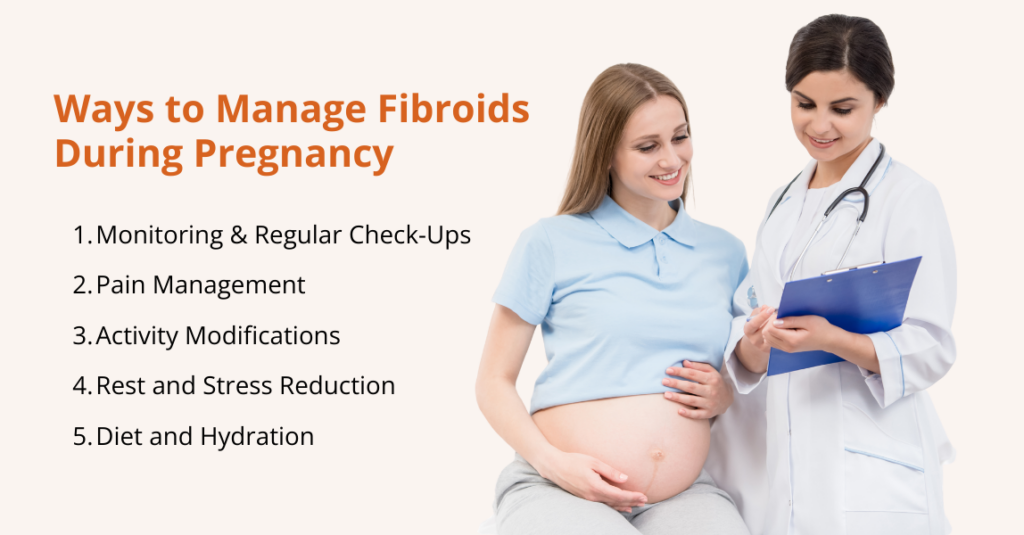
    Managing fibroids during pregnancy focuses on monitoring for complications, controlling pain, modifying activities, and maintaining overall health.

    Here are some of the most common ways to deal with fibroids during pregnancy:

    Monitoring and Regular Check-Ups

    Once fibroids are diagnosed during pregnancy, ongoing monitoring is essential to ensure both maternal and fetal health.

    Regular check-ups help track fibroid growth, assess fetal development, and identify potential complications early. Here’s how:

    • Routine Ultrasounds: These are performed periodically to track fibroid size, monitor fetal growth, and detect issues such as placental problems or cervical shortening.
    • MRI when indicated: If ultrasound images are unclear, particularly with large, multiple, or posteriorly located fibroids, MRI can provide more detailed information without radiation exposure.
    • Fetal Growth Assessments: Serial ultrasounds measure fetal size to identify intrauterine growth restriction (IUGR). If growth concerns arise, obstetricians may adjust delivery planning to ensure optimal outcomes.
    • Cervical Length Monitoring: Transvaginal ultrasounds measure cervical length, and if the cervix is short, treatments such as progesterone supplementation or cervical cerclage may be recommended.

    Regular monitoring ensures that both the mother and baby are closely observed throughout pregnancy, allowing healthcare providers to respond quickly to any complications related to fibroids.

    Pain Management

    Managing pain safely is crucial to protect both the mother and the developing baby. Treatment strategies should balance effective relief with fetal safety.

    If pain or discomfort occurs, acetaminophen (paracetamol) is one of the safest options. According to the American College of Obstetricians and Gynecologists (ACOG), when used as needed, at the lowest effective dose, and for the shortest possible time, acetaminophen remains the preferred pain and fever medicine in pregnancy.

    Untreated pain or fever itself can pose risks to a developing baby, so controlling pain wisely matters.

    Also, the U.S. Food and Drug Administration (FDA) warns that using NSAIDs after about 20 weeks can harm the baby’s kidneys, reduce the fluid around the baby (amniotic fluid), and lead to serious complications.

    Because of these risks, NSAIDs like ibuprofen or naproxen should generally be avoided after mid‑pregnancy, unless a doctor says otherwise.

    For mild pain, non‑medication options can also help. Simple measures, such as warm compresses, warm baths or showers, a pregnancy support belt, gentle stretching or prenatal yoga, and rest, can safely ease discomfort.

    Activity Modifications

    Making thoughtful adjustments to daily activities can help reduce discomfort from fibroids and support a healthy pregnancy.

    Gentle modifications, along with proper rest and nutrition, play a key role in managing symptoms safely. Here are the practical tips for activity and lifestyle:

    • Low-impact exercise: Avoid heavy lifting or high-impact workouts; choose walking, prenatal yoga, or swimming.
    • Rest and stress management: Use mindfulness, meditation, or prenatal massage to reduce discomfort.
    • Hydration and diet: Eat a high-fiber, nutrient-rich diet and stay hydrated to prevent constipation and pelvic pressure.

    These simple changes help ease symptoms and promote maternal and fetal well-being.

    Rest and Stress Reduction

    Managing stress and getting adequate rest are important for easing fibroid-related discomfort during pregnancy.

    • Relaxation Techniques: Practices such as prenatal yoga, deep breathing exercises, and mindfulness can help reduce stress and relieve pelvic or abdominal discomfort caused by fibroids.
    • Adequate Rest: Prioritizing sufficient rest allows the body to recover, lowers physical strain, and helps minimize pain, especially when fibroids contribute to pelvic pressure.

    Incorporating these habits can improve overall comfort and support a healthier pregnancy experience.

    Diet and Hydration

    Good nutrition and plenty of fluids can ease fibroid-related discomfort and support a healthy pregnancy.

    • Balanced diet: Eat regular, nutrient-rich meals with plenty of fiber, fruits, vegetables, legumes, and whole grains, plus lean protein and healthy fats. Fiber helps prevent constipation, which can worsen pelvic pressure from fibroids.
    • Staying hydrated: Drink water throughout the day to support digestion and circulation; staying well hydrated also helps reduce constipation and general discomfort.

    Together, a fiber-rich diet and consistent hydration help manage everyday stress and support overall maternal and fetal health.

    Frequently Asked Questions (FAQs)

    Do fibroids affect pregnancy?

    No. Most women with fibroids have normal pregnancies, particularly when fibroids are small or located away from the uterine cavity. The High‑Risk Pregnancy Information site estimates that only 10 %–30 % of pregnant women with fibroids develop complications. Nevertheless, because complications can be serious, women with fibroids should have enhanced prenatal care.

    Can fibroids cause pain during pregnancy?

    Yes, fibroids can cause pain during pregnancy, and it is the most common symptom associated with these growths. Pain often appears in the second or third trimester when fibroids grow faster than their blood supply, leading to a condition called red degeneration (also known as carneous degeneration). Fibroid pain may also result from mechanical pressure on nearby organs or from torsion of a pedunculated fibroid. Most mild pain can be safely managed with acetaminophen and rest. However, severe or persistent abdominal pain should prompt immediate medical evaluation, as it may indicate complications such as fibroid degeneration, placental issues, or preterm labour.

    Do fibroids grow during pregnancy?

    Yes, fibroid growth can occur during pregnancy, but patterns vary depending on the size and location of the fibroid. Many fibroids tend to grow in the first trimester due to rising estrogen and progesterone levels. Research shows that small fibroids (less than 1 cm) often increase in volume, medium-sized fibroids usually remain stable, and large fibroids (3 cm or more) may actually shrink. Interestingly, about 10–12% of fibroids regress spontaneously during pregnancy.

    Do fibroids shrink after pregnancy?

    Yes, many fibroids shrink postpartum. A case series found that fibroid size decreased in 72 % of women after giving birth and that fibroid volume could decrease by 50 % within six months. The postpartum uterus contracts to its pre‑pregnancy size, and hormonal changes (lower estrogen and progesterone levels) contribute to fibroid regression. Breastfeeding may enhance regression. However, some fibroids persist or regrow over time, so postnatal follow‑up is important.

    Conclusion

    Fibroids are common benign tumors that often coexist with pregnancy. For the majority of women, fibroids will not significantly impact fertility or pregnancy outcome.

    Nevertheless, fibroids can increase the risk of miscarriage, preterm labour, placental complications, caesarean delivery, breech presentation, and postpartum haemorrhage.

    Comprehensive prenatal care, including regular ultrasounds, growth monitoring, and blood pressure screening, allows obstetricians to detect complications early and implement appropriate interventions.

    The safest pain management strategy during pregnancy is to use acetaminophen judiciously after consulting a healthcare professional; NSAIDs should generally be avoided after 20 weeks because they can reduce amniotic fluid and harm the fetus.

    Non‑pharmacologic measures such as rest, gentle exercise, hydration, and stress reduction can help alleviate discomfort. Most fibroids regress after birth, especially with breastfeeding.

    Given this, women with fibroids should work closely with their healthcare team to develop a personalized care plan.

  • Swollen Knee: Causes, Symptoms, Treatment, & Prevention

    Swollen Knee: Causes, Symptoms, Treatment, & Prevention

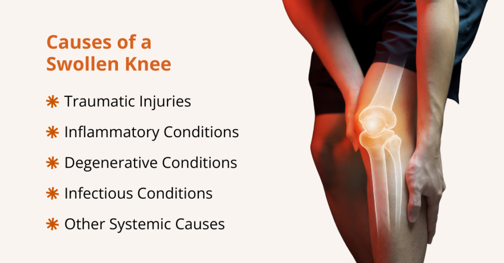
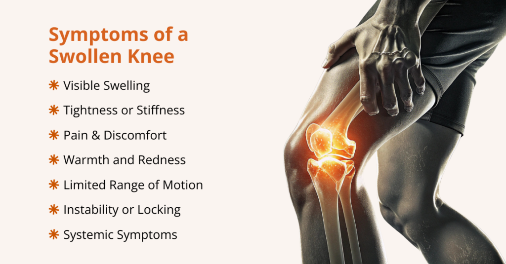
    A swollen knee, often called knee effusion, means extra fluid has collected inside the joint or in the tissues that surround it.

    Because the knee is a large hinge joint that supports your body weight and allows a wide range of motion, even mild swelling can be painful and make walking or bending difficult.

    Swelling may occur suddenly after a traumatic injury, or it can develop gradually as the result of chronic inflammation or degenerative disease.

    Although swelling itself is not a diagnosis, it is an important warning sign that something else is wrong. Causes range from minor sprains and ligament tears to more serious problems such as infections or autoimmune conditions.

    In the United States, knee injuries account for over half a million emergency department visits each year, and the lifetime prevalence of knee swelling is estimated at 27%. These numbers show how common knee problems are and why prompt attention matters.

    This guide walks through what a swollen knee is, its common causes and risk factors, symptoms to watch for, possible complications, how clinicians diagnose it, evidence-based treatment options, and practical steps you can take to reduce the risk of recurrence.

    What is a Swollen Knee?

    Knee swelling happens when extra fluid builds up in or around the knee joint. The knee connects your thigh bone (femur) to your shin bone (tibia) and kneecap (patella).

    It’s the largest joint in your body, supported by cartilage, muscles, ligaments, and nerves. Because so many structures work together, anything that irritates or injures the knee can lead to swelling.

    Swelling can occur after an injury or develop due to an underlying health condition affecting the tissues around the joint.

    When your knee is swollen, you may also notice:

    • Knee pain
    • Stiffness or a tight feeling
    • Redness or discoloration
    • Warmth or heat around the joint

    Most mild cases can be managed at home with rest, ice, elevation, and over-the-counter (OTC) medicine.

    However, you should see a healthcare provider if the swelling follows a fall or sports injury, doesn’t improve after a few days, causes severe pain, or makes it hard to move your knee.

    What Causes a Swollen Knee?

    what causes a swollen knee

    Since swelling is a symptom, not a condition in itself, it can result from a wide range of underlying conditions.

    Here are the key causes of a swollen knee:

    1. Traumatic Injuries

    Trauma is a leading cause of acute knee swelling. Injuries often occur during sports, falls, or motor vehicle collisions. When soft tissues or bone structures in the knee are damaged, blood vessels rupture, and synovial membrane irritation leads to effusion.

    These include:

    • Ligament Injuries

    Ligament tears, especially of the anterior cruciate ligament (ACL), are a leading cause of acute swelling. ACL injuries often happen with a sudden pivot or landing, and people commonly report an audible “pop,” sharp pain, rapid swelling within hours, and a feeling that the knee will give way.

    A systematic review indicates that ACL tears often accompany bone bruises, meniscus damage, or cartilage injury, all of which can lead to bleeding or joint fluid build-up (hemarthrosis), a common cause of swelling.

    Whereas posterior cruciate ligament (PCL) injuries are less common but produce similar symptoms: pain, rapid swelling, stiffness, and difficulty walking.

    • Meniscal Tears

    The menisci are crescent-shaped cartilage cushions between the femur and tibia.

    A twisting injury can tear a meniscus, causing pain, stiffness, swelling, difficulty fully straightening the knee, and occasional locking or catching.

    • Fractures

    Breaks involving the patella, distal femur, or proximal tibia can lead to substantial swelling because bone bleeding often fills the joint space (hemarthrosis).

    If the patient cannot bear weight or the leg looks deformed, prompt X-rays or CT scans are essential.

    • Dislocations

    Patellar dislocation, when the kneecap slips out of its groove, is relatively common and causes sudden pain, visible deformity, and swelling.

    2. Inflammatory Conditions

    Inflammatory arthritides can provoke episodic or chronic knee swelling. They result from autoimmune processes or crystal deposition within the synovium.

    • Rheumatoid Arthritis (RA)

    Rheumatoid arthritis is a systemic autoimmune disease that causes chronic inflammation of the synovial lining. Although it usually starts in the small joints of the hands and feet, larger joints, including the knee, are frequently affected.

    Thus, persistent synovitis causes swelling, warmth, pain, stiffness, and gradual joint damage.

    • Gout and Pseudogout (Crystal Arthropathies)

    Gout is caused by the deposition of monosodium urate crystals in joints due to hyperuricemia. While the big toe is the classic site, gout can also affect the knees, ankles, and wrists.

    Whereas Pseudogout, also called calcium pyrophosphate deposition disease (CPPD), involves deposition of calcium pyrophosphate crystals in articular cartilage and soft tissues. Flares mimic gout but are more likely to involve the knees and wrists.

    • Septic Arthritis

    Septic arthritis is an infection of the joint space, most commonly caused by bacteria. Pathogens enter the knee through hematogenous spread, direct inoculation (trauma or surgery), or extension from osteomyelitis.

    Research shows that if a knee injury with hemarthrosis is not treated promptly or properly, patients face a significantly higher risk of developing knee osteoarthritis over the next 10 to 12 years.

    The knee becomes extremely painful, swollen, warm, and difficult to move, and patients often have fever or systemic signs of infection.

    • Viral Arthritis

    Acute viral infections can cause transient arthritis due to immune activation and direct viral invasion. Common culprits include parvovirus B19, hepatitis B and C, Epstein–Barr virus, chikungunya, rubella, and arthropod‑borne viruses.

    Viral arthritis is acute, generalized inflammation of joints triggered by a broad range of viruses; it presents with joint pain, swelling, fever, rash, and fatigue, and usually resolves spontaneously once the infection is cleared.

    3. Degenerative Conditions

    Degenerative processes gradually erode joint structures, leading to chronic swelling.

    • Osteoarthritis

    Osteoarthritis is the most common cause of chronic knee swelling. It develops as cartilage breaks down and the joint becomes inflamed.

    OA affects over 32 million U.S. adults and is most common in people aged 55–64, though many younger adults are also affected.

    Typical symptoms include gradual pain that worsens with activity, stiffness after rest, creaking sensations, and occasional fluid buildup.

    • Patellofemoral Pain Syndrome (PFPS)

    Often called “runner’s knee,” PFPS causes pain at the front of the knee due to irritation in the patellofemoral joint.

    People may notice a dull ache, discomfort with running or climbing stairs, pain after sitting, and sometimes mild swelling after activity. Also, overuse, muscle imbalance, poor alignment, and improper footwear are common triggers.

    • Bursitis

    The knee contains several small bursae that reduce friction. Repetitive kneeling, irritation, or infection can inflame these sacs, leading to prepatellar or infrapatellar bursitis. Symptoms include localized swelling, tenderness, and warmth.

    4. Infectious Conditions

    In addition to septic arthritis and bursitis, infections caused by atypical organisms, such as fungi or mycobacteria, can cause joint swelling, especially in people with weakened immune systems.

    These infections may develop more slowly or present with less typical symptoms, so prompt joint aspiration, culture, and targeted (culture-directed) treatment are essential to prevent ongoing damage.

    5. Other Systemic Causes

    Some systemic diseases can lead to knee swelling through immune or metabolic processes.

    • Systemic Lupus Erythematosus (SLE)

    SLE is an autoimmune condition that can affect multiple organs, including the joints. Around 95% of patients experience joint pain or arthritis, often involving large joints like the knees.

    • Lyme Disease

    Lyme arthritis is caused by infection with Borrelia burgdorferi, transmitted via tick bites. Untreated Lyme disease often leads to recurrent knee swelling, with about 60% of cases developing arthritis.

    Most patients respond to a 30-day course of oral antibiotics like doxycycline or amoxicillin, while persistent cases may require intravenous antibiotics.

    • Hyperuricemia (Gout)

    High uric acid levels, whether from genetics, metabolic syndrome, or reduced kidney excretion, can trigger gouty flares, including in the knee.

    What are the Symptoms of a Swollen Knee?

    what are the symptoms of a swollen knee

    The signs of a swollen knee can vary depending on the cause, but common symptoms include:

    • Visible swelling around the kneecap or general enlargement of the joint.
    • Tightness or stiffness, often with a feeling of fullness or pressure.
    • Pain, ranging from mild discomfort to severe throbbing, especially when bearing weight or moving the knee.
    • Warmth and redness if inflammation or infection is present.
    • Limited range of motion, making it difficult to fully bend or straighten the knee.
    • Instability or locking, particularly with mechanical injuries like ligament or meniscus tears.
    • Systemic symptoms such as fever, chills, or fatigue, in cases of infection or autoimmune disease.

    Because swelling can hide underlying injuries or joint damage, any persistent or severe knee effusion should be evaluated by a healthcare professional.

    What are the Common Risk Factors of a Swollen Knee?

    Certain factors increase the likelihood of knee swelling. These include:

    • Age: Older adults are more susceptible to degenerative conditions, such as knee osteoarthritis.
    • Previous Knee Injuries: A history of fractures, ligament tears, or meniscal injuries predisposes to future swelling due to residual instability and cartilage damage.
    • Obesity: Excess body weight increases mechanical load on the knee, accelerating cartilage wear and predisposing to osteoarthritis.
    • Physical Activity: High‑impact sports or occupations that involve repetitive kneeling, squatting, or twisting increase injury risk. Conversely, insufficient physical activity weakens the muscles that support the knee.
    • Medical Conditions: Rheumatoid arthritis, gout, pseudogout, lupus, psoriasis, and metabolic diseases can cause recurrent joint inflammation.
    • Genetics: Family history of osteoarthritis or connective tissue disorders may increase susceptibility.

    What are the Complications of a Swollen Knee?

    what are the complications of a swollen knee

    Untreated or chronic knee effusion can lead to several complications:

    • Infection: Delayed diagnosis of septic arthritis allows bacteria to destroy cartilage and enter the bloodstream, potentially causing sepsis.
    • Chronic Pain and Disability: Persistent swelling from osteoarthritis or inflammatory arthritis results in ongoing pain, reduced mobility, and diminished quality of life.
    • Joint Instability: Ligament injuries that cause effusion may lead to ongoing instability and increased risk of recurrent injuries.
    • Reduced Range of Motion: Effusion and synovial thickening limit joint motion, leading to contractures.
    • Post‑Traumatic Osteoarthritis: Long‑standing swelling after trauma accelerates cartilage degeneration and can lead to secondary osteoarthritis.
    • Blood Clots (Deep Vein Thrombosis): Immobilization and inflammatory states increase the risk of venous thromboembolism.

    How to Diagnose a Swollen Knee?

    You can often tell a knee is swollen by comparing it to the other side: a swollen knee looks puffy or larger, the skin may appear shiny or stretched, and the joint can feel warm, tight, or painful, especially when you try to bend or straighten the leg.

    You may also notice uneven contours, reduced motion, or a sense of pressure or fullness inside the joint.

    Healthcare providers usually follow a stepwise approach to diagnose a swollen knee:

    1. Clinical Evaluation

    Diagnosing a swollen knee starts with a detailed history and physical exam:

    • History: Clinicians ask about the onset (sudden or gradual), prior injuries or infections, activity level, medications, comorbidities, and symptoms such as fever or rash.
    • Inspection & Palpation: The knee is checked for asymmetry, bruising, deformity, warmth, tenderness, effusion, and patellar mobility. Joint line tenderness is also assessed.
    • Range of Motion (ROM) & Special Tests: Active and passive ROM are compared to the other knee. Specific tests, such as the Lachman (ACL), posterior drawer (PCL), McMurray (meniscus), and patellar apprehension tests, help pinpoint injuries.

    2. Imaging Studies

    Imaging helps confirm diagnoses and assess structural damage:

    • X‑Ray: First‑line imaging to evaluate fractures, degenerative changes, and patellar alignment.
    • Ultrasound: Useful for detecting joint effusions, guiding aspirations, and evaluating bursitis or soft tissue injuries.
    • Magnetic Resonance Imaging (MRI): Gold standard for visualizing ligaments, menisci, cartilage, and bone marrow lesions. MRI can detect occult fractures, bone contusions, and synovial proliferation.
    • Computed Tomography (CT): Provides detailed bone images for complex fractures or preoperative planning.

    3. Arthrocentesis (Joint Aspiration)

    When the etiology is unclear or infection is suspected, joint aspiration is invaluable. Under sterile technique, a needle is inserted into the joint to withdraw fluid.

    The fluid is analyzed for color, clarity, viscosity, white cell count, crystals, and microorganisms. This distinguishes inflammatory from non‑inflammatory effusions, identifies septic arthritis, and diagnoses gout or pseudogout.

    Therapeutically, removing fluid can relieve pressure and improve mobility. Intra‑articular corticosteroid injection may be performed concurrently for inflammatory conditions.

    4. Laboratory Tests

    The following blood tests can aid in diagnosis:

    • Complete blood count (CBC) and C‑reactive protein (CRP)/Erythrocyte sedimentation rate (ESR)
    • Uric Acid Test
    • Rheumatoid Factor (RF) and Anti‑CCP
    • Antinuclear Antibody (ANA)
    • Culture and Gram Stain

    Swollen Knee Treatment Options

    Treatment for a swollen knee depends on the underlying cause, the severity and duration of symptoms, and other factors.

    Mild or short-term swelling from minor injuries can often be managed at home. Still, persistent, recurrent, or severe swelling may require medical evaluation and targeted interventions, or surgical options may be considered to address the issue.

    At home

    For many minor injuries and mild inflammation, conservative management suffices:

    • Rest: Avoid activities that worsen pain; short periods of limited weight‑bearing and the use of crutches may be necessary.
    • Ice: Apply cold packs for 15–20 minutes every few hours during the first 48–72 hours to reduce swelling.
    • Compression: Elastic bandages or sleeves help limit swelling; ensure they are not too tight.
    • Elevation: Keeping the knee above heart level assists venous return and reduces edema.
    • Non‑steroidal Anti‑Inflammatory Drugs (NSAIDs): Ibuprofen or naproxen relieve pain and inflammation. However, caution is needed in patients with kidney disease or gastrointestinal ulcers.
    • Activity Modification and Bracing: Temporarily avoiding high‑impact activities and using a knee brace can stabilize the joint during healing.
    • Physical Therapy: A structured rehabilitation program restores range of motion, strengthens surrounding muscles, improves balance, and reduces the risk of recurrence.

    Minimally Invasive Procedures

    When conservative measures fail, minimally invasive interventions may be considered:

    • Corticosteroid Injections: Intra‑articular corticosteroids reduce inflammation and provide rapid symptom relief for conditions like rheumatoid arthritis, gout, or OA flares. However, repeated injections may damage cartilage or increase the risk of infection.
    • Platelet‑Rich Plasma (PRP) Injections: PRP, derived from the patient’s own blood, contains growth factors that may promote tissue healing. Emerging evidence suggests benefit in early osteoarthritis and chronic tendon injuries.
    • Hyaluronic Acid (Viscosupplementation): Injecting hyaluronic acid aims to improve joint lubrication in osteoarthritis. Some patients experience modest pain relief lasting several months.
    • Cryoablation: Freezing sensory nerves can provide longer‑lasting pain relief in chronic conditions. It may be considered in select patients after other therapies fail.

    Surgical Management

    Surgery is reserved for severe injuries or advanced disease:

    • Arthroscopic Surgery: Minimally invasive arthroscopy allows surgeons to repair or remove torn meniscal fragments, trim damaged cartilage, reconstruct ligaments (ACL or PCL), or remove loose bodies.
    • Open or Arthroscopic Fracture Fixation: Stabilizes displaced fractures to facilitate healing and early mobilization.
    • Synovectomy: Surgical removal of inflamed synovial tissue may be performed in refractory rheumatoid arthritis or pigmented villonodular synovitis.
    • Total Knee Replacement (Arthroplasty): Replaces the diseased joint surfaces with artificial implants. This procedure is considered when pain and swelling severely limit function and conservative treatments fail.

    Postoperative Care and Rehabilitation

    Following surgery, appropriate postoperative care is essential:

    • Pain Management: Multimodal analgesia (NSAIDs, acetaminophen, opioids, nerve blocks) and cryotherapy help control pain.
    • Early Mobilization: Guided by surgeons and physical therapists, patients begin gentle ROM exercises and weight‑bearing as tolerated to prevent stiffness and blood clots.
    • Physiotherapy: Progressive strengthening and endurance exercises restore function and reduce the risk of complications.

    How to Prevent a Swollen Knee?

    Although not all causes of knee swelling are preventable, adopting healthy habits reduces the risk:

    • Protective Equipment: Use knee pads during sports and occupations that involve frequent kneeling or contact.
    • Weight Management: Maintaining a healthy body weight reduces mechanical stress and lowers the risk of osteoarthritis.
    • Strength Training: Strengthening quadriceps, hamstrings, gluteal, and core muscles enhances joint stability.
    • Low‑Impact Exercise: Incorporate activities like swimming, cycling, or elliptical training that minimize joint stress while maintaining cardiovascular fitness.
    • Proper Technique: Learn proper form for sports and weight lifting to avoid sudden twisting or hyperextension injuries.
    • Stretching: Regular stretching of the quadriceps, hamstrings, and calf muscles maintains flexibility and reduces strain on the knee.
    • Early Management of Chronic Diseases: Prompt treatment of autoimmune diseases, metabolic syndrome, and gout reduces flare‑ups and joint damage.
    • Regular Monitoring: Individuals at high risk (athletes, older adults, and those with prior knee injuries) should undergo periodic medical evaluations to detect early changes.

    Frequently Asked Questions (FAQs)

    Can a swollen knee be dangerous?

    Yes. While many causes are benign, rapid swelling accompanied by fever, severe pain, or inability to bear weight may signal a fracture, ligament tear, or septic arthritis and should prompt immediate medical attention. Delay in treatment increases the risk of permanent damage and systemic infection.

    What can I do for a swollen knee at home?

    Follow the RICE protocol: rest, ice, compression, and elevation. Use NSAIDs for pain if safe. Avoid activities that exacerbate symptoms. However, if swelling lasts more than a few days or recurs frequently, see a healthcare provider.

    How long does it take for knee swelling to go down?

    The time it takes for knee swelling to go down depends on the cause and severity of the injury or condition. Mild swelling from minor strains or overuse may improve within a few days to a week with rest, ice, compression, and elevation. Whereas swelling from more significant injuries, such as ligament tears, fractures, or hemarthrosis, can take several weeks to months to resolve, especially if rehabilitation or medical treatment is required. Chronic conditions such as arthritis or gout may cause intermittent swelling that persists or recurs over time and often requires ongoing management to control inflammation.

    When should I worry about a swollen knee?

    You should worry about a swollen knee if it is sudden, severe, or persistent, or comes with warning signs. Seek urgent care if you have rapid swelling, inability to bear weight, severe pain, deformity, redness, warmth, fever, or numbness. See a doctor soon if swelling doesn’t improve after a few days, recurs, causes locking or instability, or is accompanied by systemic symptoms. Early evaluation helps prevent joint damage and identify serious causes such as infection, fractures, or ligament injuries.

    What happens if a swollen knee is left untreated?

    Ignoring persistent swelling can lead to complications such as cartilage degeneration, chronic instability, decreased range of motion, and, in cases of infection, joint destruction and sepsis.

    What does a swollen knee indicate?

    A swollen knee indicates that there is excess fluid in or around the joint, which is usually a sign of an underlying problem rather than a condition on its own. It can result from injuries (ligament tears, meniscus tears, fractures), degenerative changes (osteoarthritis), infections (septic arthritis, bursitis), inflammatory or autoimmune diseases (rheumatoid arthritis, lupus), or crystal deposits (gout, pseudogout). The swelling reflects inflammation, bleeding, or fluid buildup in the knee, and its cause determines the severity, associated symptoms, and treatment required.

    What does a swollen knee look like?

    A swollen knee usually appears larger or puffier than the other knee. The skin may look shiny, stretched, or tight, and the joint can feel warm to the touch. Swelling may cause uneven contours around the kneecap or along the joint line. In some cases, redness or bruising is visible, and the knee may appear stiff or slightly deformed. Swelling can make it difficult to fully bend or straighten the leg.

    Conclusion

    A swollen knee is a symptom with many possible causes, ranging from traumatic injuries such as ACL or meniscus tears, fractures, and patellar dislocations, to inflammatory conditions such as rheumatoid arthritis, gout, and pseudogout, to degenerative osteoarthritis in older adults.

    Serious infections like septic arthritis and bursitis require prompt recognition and treatment, while less common systemic causes include SLE, Lyme disease, and metabolic disorders.

    Early diagnosis and intervention are crucial to prevent complications, reduce pain, and preserve joint function. Therefore, proper evaluation, including history, physical exam, imaging, and lab tests, helps identify the underlying cause and guide effective treatment.

  • Benign Prostatic Hyperplasia: Causes, Symptoms, & Treatments

    Benign Prostatic Hyperplasia: Causes, Symptoms, & Treatments

    Benign Prostatic Hyperplasia (BPH) is the most common prostate condition affecting men, particularly as they age.

    In the United States, over 50% of men over 50 experience symptoms of BPH, and this number rises to 90% by age 85.

    BPH is closely linked with male lower urinary tract symptoms (LUTS), but it does not raise the risk of prostate cancer or cause sexual dysfunction. However, it can affect quality of life by leading to bothersome and sometimes embarrassing urinary issues.

    While age and genetics play a key role in the development of BPH, emerging research shows that modifiable factors, including hormone levels, obesity, glucose regulation, diet, exercise, and inflammation, also influence its onset and progression.

    Therefore, understanding these risk factors, recognizing early symptoms, and seeking timely diagnosis and treatment can dramatically improve outcomes and quality of life.

    This article provides an overview of BPH, including its causes, symptoms, risk factors, diagnosis, and the latest treatment options, to help patients and caregivers understand how to manage this common condition effectively.

    What is Benign Prostatic Hyperplasia?

    what is benign prostatic hyperplasia

    BPH is the non-cancerous enlargement of the prostate gland, a small walnut-sized organ below the bladder that surrounds the urethra and produces seminal fluid.

    As men age, the prostate can enlarge from the size of a walnut to that of a lemon. In BPH, the prostate undergoes increased cell growth (hyperplasia), especially around the urethra, leading to gland enlargement.

    As it grows, it can compress the urethra and partially block urine flow, causing symptoms such as difficulty starting urination, a weak urine stream, or needing to urinate frequently.

    Who is more likely to have Benign Prostatic Hyperplasia?

    In the United States alone, BPH accounts for millions of healthcare visits each year, reflecting its prevalence among an aging male population.

    Certain risk factors make someone more prone to developing BPH:

    • Age

    Advancing age is the strongest risk factor. BPH is uncommon before 40, but the incidence rises sharply in later decades. The prostate tends to keep growing as men get older, which is why BPH primarily affects middle-aged and older men.

    • Family History (Genetics)

    Men with a family history of BPH, such as a father or brother who had an enlarged prostate, are more likely to develop the condition themselves. Studies have shown that first-degree relatives of BPH patients may have a fourfold increase in risk compared to men with no family history.

    • Diabetes and Heart Disease

    Research shows that chronic health conditions like type 2 diabetes and cardiovascular disease have been linked to a higher likelihood of BPH. Men with diabetes, in particular, have a higher incidence of BPH and often more severe symptoms, possibly due to insulin and growth-factor effects on the prostate.

    • Diet and Obesity

    An unhealthy diet and obesity can increase BPH risk. Research suggests that diets high in fat and red meat are associated with a greater risk of symptomatic BPH, whereas diets rich in vegetables may be protective. Obesity is also correlated with BPH; men who are overweight or who have metabolic syndrome tend to have larger prostate volumes and more symptoms.

    • Localized Inflammation

    Chronic prostatitis or inflammation of the prostate is often found alongside BPH on tissue analysis, and researchers suspect that inflammation may contribute to prostate enlargement. Inflammatory cells in the prostate can release factors that stimulate growth.

    • Metabolic Syndrome

    Related to the above factors, metabolic syndrome (a cluster of conditions including insulin resistance/diabetes, high blood pressure, and high cholesterol) has been linked to BPH.

    Men with metabolic syndrome tend to have larger prostates on average, and those with poor blood sugar control have an increased risk of LUTS.

    This may be due to metabolic syndrome’s association with obesity, inflammation, and hormonal imbalances that also drive prostate growth.

    • Hormone Levels

    Although not a traditional risk factor in the sense of a lifestyle choice, age-related hormonal changes underlie BPH development.

    Research shows that men who have normal testes producing testosterone can develop BPH, whereas men who were castrated or have very low testosterone from a young age do not develop BPH.

    What Causes Benign Prostatic Hyperplasia?

    what are the causes of benign prostatic hyperplasia

    Despite how common BPH is, its exact cause remains unclear. As men grow older, changes in sex hormones, particularly the accumulation of dihydrotestosterone (DHT), can stimulate prostate growth.

    Key factors contributing to BPH:

    • Aging
    • Hormonal changes
    • Family history
    • Lifestyle factors

    Other health conditions, such as obesity, type 2 diabetes, and heart and blood vessel diseases

    What are the Symptoms of Benign Prostatic Hyperplasia?

    what are the symptoms of benign prostatic hyperplasia

    BPH mainly affects urination and can range from mild to severe. As the prostate enlarges, it presses on the urethra and bladder, leading to lower urinary tract symptoms (LUTS) that can disrupt daily life.

    Common BPH symptoms include:

    • Frequent urination: Needing to urinate more often, especially at night (nocturia). Some men go every 1–2 hours.
    • Urgency: A sudden, strong urge to urinate, even when the bladder isn’t full.
    • Difficulty starting urination (hesitancy): Trouble beginning the urine stream, sometimes requiring straining.
    • Weak or interrupted urine stream: A slow or stop-and-start flow due to urethral narrowing.
    • Dribbling at the end: Small drops of urine may continue after finishing.
    • Incomplete bladder emptying: Feeling that the bladder isn’t fully emptied, leading to frequent trips to the bathroom.
    • Urinary retention: In severe cases, partial or complete blockage can make urination difficult or impossible, which is a medical emergency.
    • Painful urination (dysuria): BPH itself usually doesn’t cause burning, but infections or straining can.
    • Blood in the urine (hematuria): Straining or bladder irritation can cause small amounts of blood in the urine; a doctor should check any visible blood.

    Many of these symptoms are quantified by doctors using the International Prostate Symptom Score (IPSS), which is a questionnaire that asks about frequency, nocturia, weak stream, etc., to grade symptom severity from mild to severe.

    Also, not every man with BPH will have all these symptoms, and the severity can vary.

    What are the Complications of Benign Prostatic Hyperplasia?

    BPH is benign and usually develops slowly, but if it becomes advanced or is left untreated, it can lead to complications in the urinary tract.

    The prostate’s blockage of urine flow and the resulting strain on the bladder can cause several issues:

    • Urinary Retention: The enlarged prostate can block urine flow, causing the bladder to fill and sometimes leading to sudden, painful inability to urinate (acute retention) or constant partial retention.
    • Recurrent Urinary Tract Infections (UTIs): When the bladder doesn’t empty fully, urine can stagnate, leading to infections and symptoms such as burning, cloudy urine, or pelvic pain.
    • Bladder Stones: Minerals in leftover urine can form stones, which may worsen urinary problems or cause blood in the urine.
    • Bladder Damage: Constant pressure on the bladder can weaken its muscles and reduce its ability to empty properly. This can cause long-term urinary problems even after treatment.
    • Kidney Damage: Severe blockage can cause urine to back up into the kidneys (hydronephrosis), potentially leading to kidney injury or infection. Rarely, untreated BPH can contribute to kidney failure.
    • Incontinence: Overflow or urge incontinence can occur when the bladder is too full or unstable. Some BPH treatments may also cause temporary incontinence.
    • Blood in Urine (Hematuria): Persistent bleeding can happen due to the enlarged prostate, sometimes requiring treatment.
    • Sexual Side Effects: BPH itself doesn’t cause erectile dysfunction, but treatments may lead to issues like retrograde ejaculation, which can affect fertility but is not dangerous.

    Many of these complications are preventable with timely treatment. If you have BPH, it’s important to follow up with your healthcare provider, especially if symptoms are worsening.

    How is Benign Prostatic Hyperplasia Diagnosed?

    When BPH is suspected, healthcare providers will perform a series of evaluations to confirm the diagnosis, assess severity, and rule out other conditions (like infections or prostate cancer) that can cause similar symptoms.

    Diagnosing BPH typically involves some or all of the following steps and tests:

    • Medical history & symptom check: Your doctor will ask about urinary habits (frequency, urgency, nighttime trips, stream changes, blood in urine) and may use a questionnaire such as the IPSS/AUA symptom score to rate severity.
    • Digital rectal exam (DRE): A quick exam where the doctor feels the prostate through the rectal wall to estimate size and check for lumps or hard areas.
    • Urinalysis: A urine sample checks for infection, blood, or other conditions (e.g., diabetes) that can mimic or worsen symptoms.
    • PSA blood test: Measures prostate-specific antigen. PSA can be higher with larger prostates or cancer, so results are interpreted alongside other findings.
    • Uroflowmetry (flow test): You urinate into a device that measures flow rate and volume to see if there’s an obstruction.
    • Post-void residual (PVR) scan: A quick bladder ultrasound (or catheter measurement) shows how much urine remains after voiding—high volumes suggest incomplete emptying.
    • Transrectal ultrasound (TRUS): Used when precise prostate size is needed (for treatment planning) or before biopsy; not required in every case.
    • Cystoscopy: A scope is passed into the urethra to view the prostate and bladder directly; it is used when symptoms are unusual or before certain procedures.
    • Urodynamic testing: Specialized pressure and flow studies that evaluate bladder function; reserved for complex cases (e.g., weak bladder muscle, neurologic disease).
    • Advanced imaging (MRI/CT): Not routine for BPH, but may be used if cancer is suspected or to assess the kidneys/bladder when complications are possible.

    In most cases, a combination of history, DRE, urinalysis, PSA, and simple office tests (uroflow and PVR) is enough to diagnose BPH and start treatment.

    More invasive or specialized tests are used when the diagnosis is uncertain, symptoms are severe, or surgery is being considered.

    Benign Prostatic Hyperplasia Treatment Options

    benign prostatic hyperplasia treatment options

    Multiple treatment options exist for BPH, ranging from simple lifestyle adjustments to medications to minimally invasive procedures and surgery.

    Here we’ve outlined the main categories of BPH treatment:

    Lifestyle Changes (Conservative Management)

    For men with mild BPH symptoms, simple lifestyle adjustments can help reduce urinary problems and improve comfort:

    • Adjust fluid intake: Avoid drinking large amounts in the evening and limit fluids 1–2 hours before bedtime to reduce nighttime bathroom trips.
    • Limit bladder irritants: Cut back on caffeine, alcohol, and any foods that trigger bladder irritation, such as spicy or acidic items.
    • Stay active and maintain a healthy weight: Regular exercise (e.g., 30 minutes of brisk walking daily) can improve urinary symptoms. Maintaining a healthy weight, blood pressure, and blood sugar also supports prostate health.
    • Optimize bathroom habits: Urinate when you feel the urge, and try “double voiding” to empty the bladder more completely. Planning regular trips every 2–3 hours can help manage frequent urges.
    • Review medications: Some decongestants, antihistamines, and diuretics can worsen urinary symptoms. Consult your doctor before using these medications.
    • Pelvic floor exercises: Kegel exercises can strengthen the pelvic muscles and improve bladder control.
    • Stay warm: Cold can worsen urinary retention; dressing warmly and avoiding prolonged exposure to cold may help.

    Many men start with “watchful waiting” using these habits, often managing symptoms without medication. Regular check-ups are important to monitor progression.

    If symptoms worsen, doctors may recommend medications or other treatments.

    Medications for BPH

    For men with moderate to severe BPH, medications are usually the first line of treatment. These drugs either relax the prostate and bladder muscles to improve urine flow or shrink the prostate over time.

    Common options include:

    • Alpha-blockers

    These relax the muscles in the prostate and bladder neck, making urination easier. They work quickly (days to weeks) but do not shrink the prostate.

    • 5-Alpha-Reductase Inhibitors (5-ARIs)

    Drugs like finasteride (Proscar) and dutasteride (Avodart) shrink the prostate by blocking DHT, the hormone that drives growth. They take several months to work and are most effective in larger prostates.

    • Combination Therapy

    For men with severe symptoms or large prostates, alpha-blockers and 5-ARIs can be used together (e.g., tamsulosin + dutasteride, Jalyn®). This provides fast symptom relief and long-term prostate shrinkage.

    • Phosphodiesterase-5 inhibitors (PDE-5i)

    Tadalafil (Cialis) at a low daily dose can improve urinary symptoms by relaxing the bladder and prostate muscles. It’s especially helpful for men who also have erectile dysfunction.

    • Other Medications

    For overactive bladder symptoms, anticholinergics (e.g., oxybutynin, tolterodine) or beta-3 agonists (mirabegron) may be added. These help reduce urgency and frequency but must be used carefully if bladder emptying is poor.

    Most men start with an alpha-blocker for quick relief. If the prostate is large or symptoms persist, a 5-ARI or combination therapy may be added.

    Doctors monitor symptom scores, urine flow, and PSA levels to ensure treatment is effective and safe. If medications don’t work or side effects are unacceptable, minimally invasive procedures or surgery may be considered.

    Minimally Invasive Procedures for BPH

    For men whose symptoms aren’t fully controlled with medication or who want to avoid major surgery, minimally invasive procedures offer effective alternatives with quicker recovery and fewer complications.

    These procedures either widen the urethra or shrink prostate tissue. Common options include:

    • Prostatic Artery Embolization (PAE)

    Prostatic artery embolization is a truly minimally invasive, non-surgical procedure performed by Dr. Zagum Bhatti, an interventional radiologist.

    It involves accessing the blood vessels to the prostate (usually via a catheter inserted in the femoral or radial artery) and injecting tiny particles to block blood flow in the prostatic arteries.

    PAE is done under local anesthesia and sedation, and typically, no hospital stay is needed.

    It’s an option for men who may not tolerate surgery or anesthesia or who prefer an arterial approach. PAE’s advantages are that there’s no incision, no general anesthesia, and sexual function is generally preserved.

    • Prostatic Urethral Lift

    This is a device-based therapy where small implants are used to pull apart the enlarged prostate lobes, widening the urethral passage.

    The urologist inserts a special instrument through the urethra and places several tiny spring-like implants that tether the prostate lobes open, almost like curtain tie-backs, to prevent them from pressing inward. This does not involve cutting or destroying tissue, and it can be done under local anesthesia or light sedation.

    The benefit is that it improves urine flow and often preserves sexual function (minimal risk of retrograde ejaculation or ED) compared to traditional surgery. It is best suited for men with relatively moderate prostates and no large middle lobe.

    • Rezūm Water Vapor Therapy

    Rezūm is a newer treatment that uses steam injections to ablate prostate tissue.

    During this procedure, a device is introduced via the urethra and a small needle is deployed into the prostate; steam (water vapor) is then released, which delivers thermal energy that kills the surrounding prostate cells.

    Over the next days and weeks, the body’s immune system clears away the treated tissue, shrinking the prostate.

    • Laser Therapy

    Laser prostatectomy involves inserting a scope through the urethra and using laser energy to burn/vaporize or cut prostate tissue.

    Examples include photoselective vaporization of the prostate (PVP), often called GreenLight laser (which vaporizes tissue), and holmium laser enucleation of the prostate (HoLEP), which uses a laser to actually enucleate (carve out) the enlarged prostate tissue in lobes.

    The advantage of lasers: lower risk of bleeding, so it’s good for patients on blood thinners or with bleeding risks. However, side effects like retrograde ejaculation still occur in many cases (especially if a lot of tissue is removed).

    • Transurethral Microwave Therapy (TUMT)

    This is an older minimally invasive therapy where a special catheter with a microwave antenna is inserted into the urethra, and microwave energy heats and destroys prostate tissue from inside.

    TUMT can be done without general anesthesia. It generally only reduces symptoms moderately and may take weeks to have an effect.

    Surgical Procedures

    Modern BPH surgeries are mostly done endoscopically (through the urethra) without external cuts, providing lasting relief by removing or reducing the enlarged prostate tissue.

    Common surgical options include:

    • Simple Prostatectomy (Open or Robot-Assisted)

    Reserved for very large prostates (>80–100 grams), this involves removing the enlarged internal portion via an open incision or robot-assisted surgery.

    Recovery is longer, with a few days in the hospital and the use of a catheter. It carries higher risks like bleeding, infection, and blood clots, but offers excellent symptom relief.

    • Transurethral Incision of the Prostate (TUIP)

    TUIP is for men with small prostates (<30 cc) but significant blockage. The surgeon makes one or two small cuts in the prostate and bladder neck to widen the urethra without removing tissue.

    Recovery is usually quick, with a lower risk of retrograde ejaculation than TURP.

    In general, surgery is considered when symptoms are severe and not responding well to meds, the patient can’t tolerate meds, or if there are complications.

    How to Prevent Benign Prostatic Hyperplasia

    Since BPH is so tied to the aging process and genetics, there is no guaranteed way to prevent prostate enlargement altogether.

    Here are some strategies that may help support long-term prostate health:

    • Eat a Healthy Diet: Focus on fruits, vegetables, whole grains, and healthy fats (such as fish, nuts, and olive oil). Avoid too much red meat, high-fat foods, processed foods, sugar, and excess dairy.
    • Maintain a Healthy Weight: Obesity can disrupt hormone balance and increase the risk of BPH. Staying at a healthy weight through diet and exercise may help prevent prostate enlargement.
    • Exercise Regularly: Aim for at least 30 minutes of moderate activity, like walking, most days. Exercise improves blood flow and hormone balance and reduces inflammation, all of which support prostate health.
    • Manage Chronic Health Conditions: Keep diabetes, high blood pressure, and cholesterol under control. Good metabolic health may lower the risk of BPH progression. Avoid smoking, as it can worsen urinary symptoms.
    • Avoid Irritants: Limit caffeine and alcohol, especially in the evening, to reduce bladder stress. Stay hydrated but avoid extremes of too little or too much fluid.
    • Preventive Medications (for select cases): In certain men at higher risk (strong family history or early prostate enlargement), doctors may sometimes consider low-dose 5-alpha-reductase inhibitors to slow growth. This is not routine for all men.

    Remember, BPH is common with age, but a healthy lifestyle can delay symptoms, reduce severity, and improve well-being.

    Frequently Asked Questions (FAQs)

    How common is Benign Prostatic Hyperplasia?

    BPH is very common, especially as men get older.

    While many men may have an enlarged prostate (even without symptoms), a large portion of older men will experience BPH to some degree.

    According to the National Institute of Diabetes and Digestive and Kidney Diseases (NIDDK), BPH affects 5–6% of men aged 40–64, and 29–33% of men aged 65 and older. Similarly, Johns Hopkins Medicine estimates that about 1 in 4 men by age 55 have symptoms, and about 50% of men by age 75 do.

    Can an enlarged prostate be cured?

    An enlarged prostate can be treated with medications like 5-ARIs and procedures such as Prostatic Artery Embolization, relieving blockage and symptoms, often for many years. Since BPH is linked to aging, the prostate may grow again over time, but symptoms are usually well controlled. With proper treatment, follow-up, and healthy habits, most men regain normal urinary function and quality of life.

    What happens if you leave an enlarged prostate untreated?

    If an enlarged prostate (BPH) is left untreated, symptoms can gradually worsen and lead to serious problems: urine may not empty fully or you can suddenly be unable to urinate (acute urinary retention), recurrent urinary tract infections and bladder stones can develop, and long-term bladder strain can weaken the bladder. In severe cases, urine can back up into the kidneys (hydronephrosis) and cause kidney damage, and visible blood in the urine or new incontinence may occur. These issues reduce quality of life and can become medical emergencies.

    How long can I live with an enlarged prostate?

    You can live a long, healthy life with an enlarged prostate (BPH), as it is noncancerous and doesn’t raise the risk of prostate cancer. Life expectancy isn’t affected when symptoms are managed with lifestyle changes, medications, or procedures such as Prostatic Artery Embolization. Untreated BPH can worsen over time, but many men with mild symptoms live comfortably for years while managing them.

    What type of doctor treats benign prostatic hyperplasia?

    A urologist is the primary specialist for BPH, managing both medications and procedures such as Prostatic Artery Embolization. Primary care doctors can evaluate early symptoms and start basic treatment, but significant or persistent BPH usually requires a urologist’s care.

    Can BPH lead to prostate cancer?

    No, BPH does not cause prostate cancer. It is a benign condition, and the prostate cells are not cancerous. BPH does not raise your risk of developing prostate cancer. However, because both conditions can cause similar symptoms, such as frequent or difficult urination, regular checkups are important to rule out cancer.

    Conclusion

    Benign Prostatic Hyperplasia is a very common and manageable condition that affects a great number of aging men.

    While the prospect of an “enlarged prostate” can be worrisome, it’s important to remember that BPH is benign, it is not prostate cancer, and in itself is not life-threatening.

    The primary impact of BPH is on quality of life: it can cause frustrating urinary symptoms that interrupt sleep, work, and daily comfort.

    With modern medications and minimally invasive treatments, most men can find relief that suits their needs and lifestyle.

    If you’re a man over 50 experiencing urinary changes, such as increased frequency, hesitancy, or nocturia, it’s important to talk to your healthcare provider.

    That said, a simple evaluation can identify BPH as the cause and guide you to the most appropriate treatment plan.

    Today, men don’t have to live with severe urinary problems like previous generations. Early consultation and personalized care can keep BPH a minor concern rather than a major obstacle to healthy aging.

  • Knee Osteoarthritis: Causes, Symptoms, Diagnosis, Treatment, & Prevention

    Knee Osteoarthritis: Causes, Symptoms, Diagnosis, Treatment, & Prevention

    Knee osteoarthritis (OA) is a common and growing public health concern in the United States.

    As people live longer and rates of obesity rise, more Americans are developing this degenerative joint disease, leading to major personal and societal costs.

    In fact, research estimates that about 14 million U.S. adults suffer from symptomatic knee OA.

    Moreover, radiographic studies from the U.S. show that more than one in three adults aged 60 or older have signs of knee OA on imaging, and around 12 percent report symptoms.

    As the population ages and obesity continues to climb, resulting in knee OA prevalence rising further, understanding risk factors, diagnosis, and treatment is more important than ever.

    In this article, we’ll walk through what knee osteoarthritis is, how it progresses, who’s most at risk, what causes it, how it’s diagnosed, treatment options, and finally, strategies for prevention.

    What is Knee Osteoarthritis?

    what is knee osteoarthritis

    Knee osteoarthritis occurs when the cartilage that cushions your knee joint gradually wears down. As this protective layer thins, the bones begin to rub against each other, creating friction that leads to pain, swelling, and stiffness.

    Because the knee carries your body weight and absorbs impact with every step, it’s one of the joints most commonly affected by this degenerative “wear-and-tear” disease.

    Research shows that OA of the knee joint is often associated with aging, but it can also be influenced by factors such as injury, obesity, genetics, and other health conditions.

    As the condition worsens, individuals may experience difficulty performing daily activities, such as walking, climbing stairs, or getting up from a chair. Over time, though, the disease can worsen and may eventually limit mobility.

    However, many treatments can help slow its progression, reduce pain, and improve everyday function. Healthcare providers carefully monitor its progression, and if knee OA begins to significantly affect your quality of life, surgical and non-surgical options are available.

    What are the Stages of Knee Osteoarthritis?

    what are the stages of knee osteoarthritis

    Although there’s no single universal staging system, knee osteoarthritis typically follows a predictable, gradual course.

    Clinicians often describe it in stages to explain how advanced the disease is and what to expect.

    Knowing these stages can help you recognize symptoms early and plan treatment with your healthcare team.

    • Stage 1 (Minor)

    At this earliest stage, there is minor wear and tear to the cartilage. Changes are usually subtle, and most people feel little or no pain. X-rays may show only very subtle changes, if any at all.

    • Stage 2 (Mild)

    Mild OA means the cartilage is beginning to break down. You may notice occasional pain or stiffness, especially after activity or when getting up from a seated position. There is still enough cartilage to prevent bones from rubbing together, but symptoms are starting to appear.

    • Stage 3 (Moderate)

    In the moderate stage, cartilage loss is more pronounced. Pain becomes more frequent and may interfere with daily activities such as walking, climbing stairs, squatting, or kneeling. Stiffness and reduced range of motion are common, and you may feel discomfort after periods of rest.

    • Stage 4 (Severe)

    Severe OA indicates that cartilage is nearly or completely gone in parts of the knee. Bones may grind together, causing constant pain, swelling, marked stiffness, and significant limitations in movement. At this point, conservative measures often provide limited relief, and joint replacement surgery may be discussed as an option.

    What Causes Knee Osteoarthritis?

    what are the causes of knee osteoarthritis

    Knee osteoarthritis is caused by the gradual breakdown of cartilage in the knee joint.

    While the exact cause of this breakdown is not always clear, several factors contribute to its development:

    • Mechanical Stress: Research shows that overuse of the knee joint, especially among individuals who engage in repetitive activities or heavy lifting, can cause cartilage wear and tear.
    • Inflammation: Chronic joint inflammation, often due to conditions such as rheumatoid arthritis, can accelerate cartilage breakdown.
    • Genetics: Modern research shows genetic factors can contribute to the development of knee osteoarthritis. If you have a family history of the condition, you may be more likely to develop it yourself.
    • Joint Instability: Knee instability due to prior injuries or muscle strength imbalances can place additional stress on the joint, leading to cartilage damage.
    • Obesity: Studies show that excess body weight puts additional pressure on the knee joint, leading to increased cartilage wear and tear.

    What are the Symptoms of Knee Osteoarthritis?

    The symptoms of knee osteoarthritis can vary depending on the severity of the condition.

    Knee pain is the most common and noticeable symptom of knee osteoarthritis. You may feel discomfort when putting weight on your knee, during movement, or even while resting.

    As the condition progresses, other symptoms can appear, including:

    • Stiffness, especially when you first wake up or after sitting for long periods.
    • Swelling or a puffy feeling around the knee joint.
    • Cracking, popping, or grinding sounds (called crepitus) when you move the knee.
    • A feeling of instability, as if the knee might buckle or “give out.”
    • Locking or catching, where the knee briefly feels stuck or difficult to move.

    These symptoms may come and go at first, but often become more frequent over time as osteoarthritis progresses.

    Who is at Risk for Knee Osteoarthritis?

    Several factors can increase the likelihood of developing knee osteoarthritis. Some of the key risk factors include:

    • Age: As we age, the cartilage in our joints naturally wears down, increasing the risk of developing OA. It is most common in people over 50.
    • Gender: Women are more likely than men to develop knee osteoarthritis, especially after menopause. This may be due to hormonal changes that affect the joints.
    • Obesity: Excess body weight adds stress to the knee joint, increasing the risk of cartilage wear. Studies show that individuals with a body mass index (BMI) of 30 or higher are at significantly higher risk of developing knee OA.
    • Previous Joint Injury: Individuals who have experienced a knee injury, such as a ligament tear or fracture, are at higher risk for developing OA later in life. Even if the injury heals, the knee joint may be weakened, leading to cartilage damage.
    • Genetics: Some people may have a genetic predisposition to osteoarthritis. Certain genes may make individuals more susceptible to cartilage breakdown in the knee joint.
    • Occupation: Jobs that require heavy lifting, kneeling, or squatting can increase the risk of developing knee osteoarthritis due to repetitive stress on the joint.
    • Other Health Conditions: Conditions such as rheumatoid arthritis, gout, and diabetes can also increase the likelihood of developing knee osteoarthritis.

    How is Knee Osteoarthritis Diagnosed?

    how is knee osteoarthritis diagnosed

    Knee osteoarthritis is typically diagnosed through a combination of physical examination, medical history, and imaging.

    The following steps may be involved:

    • Physical Examination: Your doctor will assess your knee’s range of motion, check for swelling or tenderness, and evaluate how well your knee functions during movement.
    • Medical History: Your doctor will ask about your symptoms, any previous knee injuries, and your family history of osteoarthritis or other joint disorders.
    • X-rays: X-rays are commonly used to evaluate the extent of joint damage and cartilage loss. They can show narrowing of the joint space, bone spurs, and other signs of OA.
    • MRI: An MRI may be used if your doctor needs more detailed images of the soft tissues in your knee, including cartilage, ligaments, and tendons.
    • Blood Tests: In some cases, blood tests may be done to rule out other conditions, such as rheumatoid arthritis, that may cause similar symptoms.

    Knee Osteoarthritis Treatment Options

    Knee osteoarthritis treatment focuses on relieving pain, improving mobility, and slowing the progression of joint damage.

    Options range from lifestyle changes and nonsurgical therapies to surgical procedures. In most cases, healthcare providers begin with conservative (nonsurgical) treatments before considering surgery.

    Nonsurgical treatments for knee OA

    Nonsurgical options aim to reduce pain and inflammation, support joint function, and delay the need for surgery. Common treatments include:

    • Pain Medications: Over-the-counter NSAIDs (such as ibuprofen) or acetaminophen can help reduce pain and swelling. Prescription options may be used if symptoms are more severe.
    • Physical therapy: Targeted exercises strengthen the muscles around the knee, improve joint stability, and increase flexibility, making daily activities easier.
    • Weight loss (when needed): Reducing body weight decreases stress on the knee joint. Even losing 5–10% of body weight can significantly reduce symptoms.
    • Knee braces: Braces help support the joint, reduce strain, and may shift pressure away from the most damaged part of the knee.
    • Corticosteroid (steroid) injections: These injections reduce inflammation and can provide short-term pain relief, especially during flare-ups.
    • Knee gel injections (viscosupplementation): Hyaluronic acid “gel shots” add lubrication inside the joint to improve movement and reduce pain.
    • Genicular nerve block injections: These target the small nerves around the knee to interrupt pain signals temporarily.
    • Platelet-rich plasma (PRP) injections: PRP uses concentrated platelets from your own blood to promote healing and reduce joint inflammation.
    • Genicular artery embolization (GAE): A minimally invasive procedure that reduces blood flow to inflamed tissues around the knee, helping decrease pain and swelling for some patients.

    Surgical treatments for knee OA

    Surgery is typically considered when pain is severe, daily function is limited, or nonsurgical treatments are no longer effective. Surgical options include:

    • Cartilage restoration or replacement: Techniques such as microfracture, grafting, or implanting new cartilage aim to repair small areas of damage in younger, active patients.
    • Knee bone reshaping (osteotomy): Surgeons cut and realign the bones around the knee to shift weight away from the damaged area, helping relieve pain and delay joint replacement.
    • Partial knee replacement: Only the damaged portion of the knee is replaced with an artificial implant. This option preserves more of your natural knee and may lead to a faster recovery.
    • Total knee replacement: The entire knee joint is replaced with artificial components. This is the most common surgical treatment for advanced knee OA and can provide long-lasting pain relief and improved mobility.

    How Can Knee Osteoarthritis Be Prevented?

    While you cannot entirely prevent knee osteoarthritis, there are several steps you can take to reduce your risk and protect your knee joint:

    • Exercise Regularly: Engaging in low-impact activities like swimming, cycling, and walking can help strengthen the muscles around the knee and improve joint stability.
    • Maintain a Healthy Weight: Reducing excess weight will reduce the pressure on your knees and help protect against OA.
    • Avoid Joint Injuries: Protect your knees during physical activities, especially high-impact sports, by using proper techniques and wearing protective gear.
    • Strengthen Your Muscles: Strengthening the muscles around the knee joint can help support the joint and reduce the risk of developing osteoarthritis.
    • Stretch Regularly: Regular stretching can improve flexibility and reduce stiffness in the knee joint.

    With these prevention strategies and staying proactive about your knee health, you can reduce your risk of developing knee osteoarthritis and maintain a high quality of life for years to come.

    Frequently Asked Questions (FAQs)

    What does knee osteoarthritis feel like?

    Knee osteoarthritis often feels like a combination of pain, stiffness, and joint grinding. You may notice aching pain during movement or after activity, stiffness when you first wake up or stand after sitting, and a cracking or grating sensation (crepitus) when the knee moves. Many people also experience knee swelling, a feeling that the knee is unstable or might “give out,” and occasional locking or catching when the joint gets stuck. As the condition progresses, you may find it harder to fully bend or straighten your knee, making everyday activities more difficult.

  • Urinary Incontinence: Types, Causes, Symptoms, & Treatments

    Urinary Incontinence: Types, Causes, Symptoms, & Treatments

    Urinary incontinence (UI) means leaking urine when you don’t want to. It is a common health problem that affects millions of people, especially women and older adults.

    Over 33 million Americans suffer from some type of urinary incontinence or bladder condition.

    Although it becomes more common with age, it is not a normal part of getting older. UI usually happens because the bladder cannot store urine properly, or the muscles that hold urine in become weak.

    Importantly, it can affect daily life. People may feel embarrassed, avoid social activities, or feel stressed. For caregivers and family, UI increases physical and emotional strain.

    But do you know what makes this problem even harder?

    There is a lot of stigma around urinary incontinence, which makes people think leaking urine is “normal” after childbirth or as they get older. This stops many from asking for help.

    In this blog, we’ll break down everything you need to know about urinary incontinence, its types, causes, symptoms, risks, diagnosis, treatment, and prevention.

    With the right information, people can get help sooner, and healthcare providers can offer better care.

    What is Urinary Incontinence?

    Urinary incontinence (UI) means losing control of your bladder and accidentally leaking urine.

    This can happen in different ways: you might leak a little when you cough or sneeze, feel a sudden urge to pee, or, in rare cases, lose full control of your bladder.

    Your urinary system includes several organs that work together to filter, store, and remove waste as urine. When everything is working normally, you can get to the bathroom on time.

    Incontinence happens when these organs or muscles don’t function properly. There are many reasons this can occur, and it can affect people at any stage of life.

    While it’s true that the risk increases with age, UI can affect anyone, young or old.

    The good news is that treatments are available to help manage it. With the right care, incontinence doesn’t have to disrupt your life or stop you from staying active.

    Types of Urinary Incontinence

    types of urinary incontinence

    There are several types of urinary incontinence, each with its own causes, symptoms, and triggers. Understanding which type you have is an important step in getting the right treatment.

    The main types of incontinence include:

    1. Stress Incontinence (SUI)

    Stress incontinence (SUI) occurs when the pelvic floor muscles and/or urethral sphincter cannot resist sudden increases in intra‑abdominal pressure. Activities such as coughing, sneezing, laughing, exercising, or lifting heavy objects cause involuntary leakage.

    In women, SUI commonly results from pregnancy, vaginal deliveries, and menopause, which weaken pelvic supports and the urethra. In men, it is frequently a postoperative complication of radical prostatectomy or transurethral resection of the prostate.

    SUI is the most prevalent type in women; a cross‑sectional analysis of U.S. adults found that stress incontinence occurred in about 37.5% of women reporting incontinence.

    2. Urge Incontinence (UUI)

    Urge incontinence (UUI) is characterized by a sudden, intense urge to urinate followed by uncontrollable leakage. It is commonly associated with overactive bladder (OAB), a syndrome of urinary frequency, urgency, and nocturia.

    Detrusor muscle overactivity is the principal mechanism; triggers include bladder inflammation or irritation (such as urinary tract infections), neurologic disorders (e.g., stroke, multiple sclerosis, Parkinson’s disease), and aging.

    According to NHANES data, UUI affects approximately 9–31% of U.S. women and 2.6–21% of men, with prevalence rising sharply after age 75.

    3. Mixed Incontinence (MUI)

    Mixed incontinence (MUI) combines both stress and urgency symptoms. Research shows that 20–30% of individuals with chronic incontinence have MUI.

    People may experience leakage with physical activity and a sudden urge to void. It is common in older women and is associated with the same risk factors as SUI and UUI.

    4. Overflow Incontinence (OFI)

    Overflow incontinence results from chronic urinary retention; the bladder becomes overdistended and leaks constantly or intermittently.

    Causes include obstruction of urine outflow (e.g., enlarged prostate, urethral stricture), neurologic diseases causing impaired detrusor contractility (e.g., diabetic neuropathy, spinal cord injury), or medications that affect bladder emptying.

    Also, overflow incontinence is potentially dangerous because it can lead to urinary tract infections and, in severe cases, kidney damage.

    5. Functional Incontinence (FUI)

    Functional incontinence arises when a person cannot reach the toilet or remove clothing in time.

    Causes are external to the urinary tract, mobility impairments, cognitive disorders such as dementia, visual impairment, or environmental barriers.

    While often overlooked, functional incontinence significantly contributes to incontinence in frail older adults and nursing home residents.

    What Causes Urinary Incontinence?

    what causes urinary incontinence

    Urinary Incontinence has multifactorial causes that can be temporary or persistent. Recognising the underlying cause is essential because treatment strategies vary.

    Temporary Causes

    • Urinary Tract Infections (UTIs): Infections can irritate the bladder, leading to sudden urges to urinate and leakage.
    • Dietary Triggers: Foods and drinks like caffeine, alcohol, spicy foods, citrus fruits, carbonated drinks, and artificial sweeteners can irritate the bladder. Drinking large amounts of fluid or taking diuretics can also overwhelm the bladder.
    • Medications: Diuretics increase urine production, while some sedatives, muscle relaxants, or anticholinesterase drugs can interfere with bladder or sphincter control.
    • Constipation: Hard or impacted stool can put pressure on the bladder and block urine flow.
    • Acute Illnesses or Delirium: Confusion from illness or delirium can make it harder to recognize the need to use the bathroom or get there in time.

    Persistent or Long-Term Causes

    Key persistent and long-term causes include:

    • Pregnancy, Childbirth, and Menopause

    Pregnancy and childbirth stretch and injure pelvic tissues and nerves. Vaginal delivery, instrument‑assisted birth, and having multiple births increase the risk of later SUI.

    During menopause, declining estrogen causes atrophy of the urethral mucosa and pelvic connective tissue, reducing urethral closure pressure.

    • Aging

    Age‑related changes include reduced bladder capacity, diminished urethral sphincter tone, and decreased estrogen in women.

    Detrusor muscle overactivity becomes more frequent with aging. Evidence from the CDC’s Rise for Health study shows that women with multiple chronic conditions had poorer bladder health than those with zero or one chronic condition.

    • Overweight and Obesity

    Excess body weight increases intra‑abdominal pressure and weakens pelvic floor muscles.

    Another cross‑sectional study from NHANES 2013‑2018 reported that the weight‑adjusted waist index (WWI) was positively associated with urge urinary incontinence; each unit increase in WWI was associated with a 20% increase in UUI risk.

    The study concluded that weight‑loss interventions could reduce UI in overweight women and clinically obese men.

    • Neurological Disorders

    Stroke, spinal cord injury, Parkinson’s disease, multiple sclerosis (MS), and diabetic neuropathy can disrupt neural control of the bladder and urethra.

    For example, MS and spinal cord injury can cause detrusor overactivity or detrusor-sphincter dyssynergia (outflow obstruction). Alzheimer’s disease and other dementias contribute to functional incontinence by impairing recognition of bladder signals or the ability to reach a toilet.

    • Pelvic Floor Disorders and Connective Tissue Weakness

    Pelvic organ prolapse, such as cystocele or rectocele, can displace the bladder and urethra. Loss of connective tissue strength (e.g., collagen disorders) predisposes to SUI. In men, benign prostatic hyperplasia (BPH) and prostate cancer can cause obstruction and overflow or stress UI.

    • Diseases and Comorbidities

    Diabetes, chronic obstructive pulmonary disease (COPD), chronic kidney disease, asthma, and cardiovascular disease contribute to UI risk. Obesity‑related metabolic syndrome amplifies risk through systemic inflammation and hormonal changes.

    A study using NHANES data found that sarcopenia was independently associated with increased risk of mixed and stress UI among women aged ≥60 and that sarcopenic obesity with a metabolically unhealthy phenotype conferred the highest risk.

    What are the Symptoms of Urinary Incontinence

    symptoms of urinary incontinence

    The main symptom of urinary incontinence is leaking urine before reaching the bathroom or during activities. Leaks can be small or large, occasional or constant, and the exact symptoms often depend on the type of incontinence.

    Common symptoms may include:

    • Leaking urine during activities like coughing, sneezing, laughing, exercising, bending, or sexual activity
    • Bedwetting (enuresis)
    • Sudden, strong urge to urinate followed by involuntary leakage
    • Feeling that the bladder is full or unable to completely empty
    • Weak urine stream or needing to strain to urinate
    • Continuous dribbling or leakage without warning
    • Difficulty reaching the toilet in time due to mobility or cognitive issues

    Who Is More Likely to Develop Urinary Incontinence?

    Women are roughly twice as likely as men to experience UI; hormonal changes, pregnancy, and childbirth account for much of this difference.

    Moreover, age is a strong predictor; prevalence increases from 3.7% among people aged 65–69 to 10.6% among those aged ≥85.

    Race/ethnicity and socioeconomic status also influence risk; African American women have higher rates of urge or mixed UI, while white women are more likely to report stress UI.

    Also, higher body‑mass index, diabetes, COPD, hypertension, and neurological disorders increase risk. Similarly, a study shows that sarcopenia, sarcopenic obesity, and metabolic unhealthy obesity were shown to elevate the risk of stress and mixed UI markedly.

    In addition, research also highlights that environmental exposures to endocrine‑disrupting chemicals (e.g., bisphenol A) may specifically raise the risk of urge UI.

    What are the Complications of Urinary Incontinence?

    what are the complications of urinary incontinence

    Chronic UI can lead to:

    • Dermatologic Problems: Persistent wetness causes skin irritation, rashes, infections, and pressure ulcers.
    • Urinary Tract Infections (UTIs): Incomplete emptying and catheter use increase the risk of UTIs.
    • Reduced Quality of Life: People may restrict social interactions and physical activities to avoid accidents, leading to isolation, depression, and anxiety.
    • Falls and Fractures: Rushing to the toilet increases the risk of falls, particularly among older adults.
    • Sleep Disruption: Nocturnal urgency and voiding disturb sleep, causing fatigue and cognitive impairment.
    • Caregiver Burden and Institutionalization: UI is a major reason for admission to long‑term care, and study shows that more than 50% of nursing home residents and 75% of long‑term care residents have UI.
    • Kidney Damage: Chronic urinary retention in overflow incontinence can lead to hydronephrosis and renal failure.

    How Is Urinary Incontinence Diagnosed?

    A thorough evaluation is essential to determine the type and cause of UI. Here’s how:

    • History & Physical Examination

    The Doctor asks about when and how often leaks happen, fluid intake, medications, and health history. They check your abdomen/pelvis and may do a “cough stress test” to see if leakage happens with pressure.

    • Bladder Diary

    You record when you drink, when you pee, how much, and when leaks occur (for several days). This helps identify patterns and triggers.

    • Urinalysis & (if needed) Urine Culture

    A urinalysis checks for urinary tract infection (UTI), blood, sugar, or other abnormalities that might explain incontinence. Also, blood tests are sometimes performed to evaluate kidney function or detect other systemic conditions. These blood tests assess renal function, glucose, calcium, and electrolytes.

    • Post-Void Residual Measurement

    After you urinate, a test (via ultrasound or catheter) may measure how much urine remains in your bladder. If a large amount remains, this may indicate incomplete bladder emptying (overflow or neurogenic bladder).

    • Bladder Function (Urodynamic) Tests

    For more complex or unclear cases, tests such as uroflowmetry, cystometry, or pressure-flow studies assess how well your bladder and sphincter store and release urine.

    • Cystoscopy or Imaging

    If there’s suspicion, endoscopic or radiographic evaluation is performed to assess for abnormalities, bladder stones, tumors, or fistulas.

    Urinary Incontinence Treatment Options

    urinary incontinence treatment options

    Treatment should be individualized based on the type of incontinence, severity, age, sex, and comorbidities.

    Management usually follows a stepwise approach: lifestyle modifications, behavioral therapies, medications, devices, and surgery.

    Lifestyle Changes

    These are first‑line strategies recommended for all patients because they have minimal side effects and address reversible factors.

    A frontiers study of 20,195 adults found that adherence to 4–5 healthy lifestyle factors (non‑smoking, moderate alcohol intake, regular physical activity, healthy diet, and optimal waist circumference) reduced the risk of overactive bladder by 46% compared with adherence to 0–1 factors.

    Here are some lifestyle and behavioral therapies to consider:

    • Pelvic Floor Muscle Training (PFMT)

    Also called Kegel exercises, PFMT strengthens the levator ani and urethral sphincter. People contract and relax pelvic muscles in sets of 10–15 squeezes, three times daily.

    Evidence indicates that PFMT improves or resolves symptoms in two-thirds of women. Men benefit as well; PFMT is recommended after prostate surgery.

    • Lifestyle Modifications

    Maintain a healthy weight, treat chronic cough, manage diabetes and constipation, stop smoking, reduce alcohol and caffeine intake, and avoid bladder irritants.

    • Fluid Management

    Drink adequate fluids (about 6–8 glasses daily) and avoid excessive intake. Avoid drinking right before bedtime.

    Behavioral Therapies

    Behavioral therapies help retrain the brain–bladder connection and reduce anxiety, urgency, and leakage through mental and emotional techniques.

    • Bladder Training and Timed Voiding

    For urgency or mixed incontinence, patients gradually increase intervals between voids and use urge‑suppression techniques. This helps expand bladder capacity and reduce urgency.

    • Cognitive Behavioral Therapy (CBT)

    CBT helps individuals modify thoughts and behaviors that exacerbate urgency or fear of leaking. It teaches coping strategies, reduces bathroom-related anxiety, and improves confidence in bladder control.

    Physical Therapies

    Physical therapy focuses on strengthening and retraining the pelvic floor muscles to improve bladder control and reduce leakage.

    • Pelvic Floor Muscle Rehabilitation

    This involves learning to strengthen and control the pelvic floor muscles, as they support the bladder and help prevent urinary leakage.

    A physical therapist teaches proper techniques (similar to Kegel exercises). Therapy may also include breathing techniques and core strengthening to improve overall pelvic stability.

    • Biofeedback and Electrical Stimulation

    Biofeedback uses sensors to display muscle activity on a screen, helping you learn when you’re contracting the right muscles and how to improve control.

    Whereas electrical stimulation delivers a gentle current to activate weak pelvic floor muscles, strengthen them over time, and reduce symptoms like urgency, frequency, and leakage.

    Together, they help improve muscle awareness, coordination, and bladder control.

    Medications

    Medication is usually considered when behavioral therapies don’t provide enough relief.

    Common drug options include:

    • Anticholinergics (Antimuscarinics)

    Antimuscarinic agents (e.g., oxybutynin, tolterodine, solifenacin) are used to relax the bladder muscle to reduce urgency, frequency, and urge-related leakage.

    In U.S. Medicare data, antimuscarinics remain the most commonly prescribed, but their use decreased from 49% to 34% between 2012 and 2021, while β‑3 agonist use increased from 0.2% to 17%.

    • Beta-3 Agonists

    These drugs (e.g., mirabegron, vibegron) also relax the bladder muscle but typically have fewer cognitive side effects than anticholinergics.

    • Topical Estrogen

    Low-dose vaginal estrogen can improve urethral and vaginal tissue health, reduce irritation, and improve stress or urgency symptoms in postmenopausal women.

    It is not the same as systemic hormone therapy and has minimal systemic absorption.

    • Medications for Men with BPH-Related Incontinence

    For men with bladder symptoms caused by prostate enlargement, alpha-blockers (e.g., tamsulosin, terazosin) help relax the prostate and bladder neck. Whereas 5-alpha reductase inhibitors (e.g., finasteride, dutasteride) shrink the prostate over time. Often, these medications are combined for better symptom control.

    Minimally Invasive Procedures

    These treatments are considered when lifestyle changes and medications aren’t enough and offer effective, low-risk options to improve bladder control.

    • Botox Injections for Overactive Bladder

    Botox is injected into the bladder muscle to calm overactive contractions. This reduces urgency, frequency, and the risk of sudden leakage. Results typically last 6–12 months.

    • Sacral Neuromodulation (Nerve Stimulation Therapy)

    A small device sends gentle electrical pulses to the sacral nerves, which control bladder function. This helps restore normal signaling and reduces urge incontinence and urinary retention.

    • Urethral Bulking Agents

    A gel-like material is injected around the urethra to facilitate closure. This provides extra support and reduces stress incontinence, especially in women with weak sphincter muscles.

    Surgical Treatments

    These procedures are considered when other treatments fail or when incontinence is severe and linked to structural problems.

    • Mid‑urethral Sling Procedures

    A mesh or tissue sling is placed under the urethra to provide support. It helps maintain urethral closure during coughing, laughing, or exercise, making it highly effective for stress urinary incontinence in women.

    • Artificial Urinary Sphincter (AUS) Implantation

    AUS is most commonly used in men, especially after prostate surgery. It involves placing an inflatable cuff around the urethra, which opens and closes via a small pump. This provides strong control for moderate to severe incontinence.

    • Bladder Neck Suspension

    This surgery lifts and secures the bladder neck and urethra into a better position. It helps reduce leakage caused by weak support tissues and is often used for stress incontinence in women.

    • Cystoplasty (Bladder Augmentation)

    This procedure enlarges the bladder using a piece of bowel. It increases bladder capacity and reduces pressure, making it useful for severe urge incontinence or neurogenic bladder when other treatments have failed.

    How to Prevent Urinary Incontinence?

    how to prevent urinary incontinence

    You can lower your risk of urinary incontinence by protecting your pelvic floor and managing lifestyle factors: Here’s how:

    • Maintain a Healthy Weight: Keeping your BMI in a healthy range reduces pressure on your bladder. Weight-loss programs are especially effective for overweight women and obese men.
    • Avoid Bladder Irritants: Cut back on caffeine, alcohol, carbonated drinks, artificial sweeteners, spicy foods, and citrus. Some people also react to chocolate and acidic foods.
    • Stay Hydrated: Drink enough water to keep urine light-colored, but don’t overdrink. Dehydration can irritate the bladder and increase the risk of UTIs.
    • Prevent Constipation: Eat more fiber, drink plenty of fluids, and stay active. Constipation can worsen bladder leakage.
    • Do Pelvic Floor Exercises: Practice PFMT regularly, including during and after pregnancy, to keep pelvic muscles strong.
    • Quit Smoking: Smoking causes chronic coughing, which strains pelvic muscles and increases SUI risk.
    • Manage Chronic Conditions: Keep conditions like diabetes, hypertension, asthma, and COPD under control to protect bladder function.
    • Stay Physically Active: Regular exercise supports weight control and muscle strength. Avoid too many high-impact activities if they trigger leakage; balance them with PFMT.

    Frequently Asked Questions (FAQs)

    Can urinary incontinence be cured?

    Many people achieve significant symptom improvement or complete resolution, especially with early intervention. Lifestyle and behavioral therapies (PFMT, bladder training) are effective first‑line treatments. For persistent symptoms, medications, neuromodulation, or surgery can provide relief. Cure rates vary by type and severity; for example, research shows that PFMT cures or improves symptoms in roughly 67% of women, while mid‑urethral sling surgery for SUI has long‑term success rates around 80%. Urge incontinence often requires combination therapy; Botox and sacral neuromodulation have similar efficacy at two years.

    Is urinary incontinence a normal part of aging?

    No. Although prevalence increases with age, UI is a medical condition, not an inevitable consequence of aging. Many older adults maintain continence with proper bladder health habits, pelvic floor exercises, and management of chronic diseases.

    Will drinking less water help with incontinence?

    Restricting fluid intake can worsen urinary symptoms because concentrated urine irritates the bladder. Instead, spread fluid intake throughout the day and limit fluids before bedtime. Avoid caffeinated or carbonated beverages, as they can’t stimulate the bladder.

    When should I see a healthcare provider about incontinence?

    You should consult a clinician if you experience involuntary leakage that affects daily life; have associated symptoms such as burning, pain, blood in urine, or frequent UTIs; or have difficulty emptying your bladder. Early evaluation helps identify reversible causes and prevents complications.

    Conclusion

    For many people, talking about bathroom habits can feel embarrassing. However, it is a common and often overlooked condition that affects millions of Americans.

    Urinary incontinence may make you self-conscious or keep you from enjoying daily activities because you’re worried about leaking or not reaching the bathroom in time.

    Therefore, promoting bladder health, encouraging early symptom reporting, and supporting lifestyle changes are essential.

    Most individuals can achieve meaningful improvement through weight management, pelvic floor exercises, bladder training, and appropriate medications or procedures.

    If you’re experiencing any signs of incontinence, don’t wait; taking action now can help you prevent discomfort and bigger problems later in life.