One of the most important things to understand about uterine fibroid embolization before you decide to have the procedure is what the recovery actually looks like. UFE is often described as minimally invasive — which it is, compared to hysterectomy or myomectomy — but it does have a recovery period that requires preparation and realistic expectations.
The post-UFE experience divides naturally into phases. Understanding what is normal at each phase, what to watch for, and when you can expect to resume your normal activities helps you plan practically and reduces anxiety about symptoms that are actually part of the expected healing process.
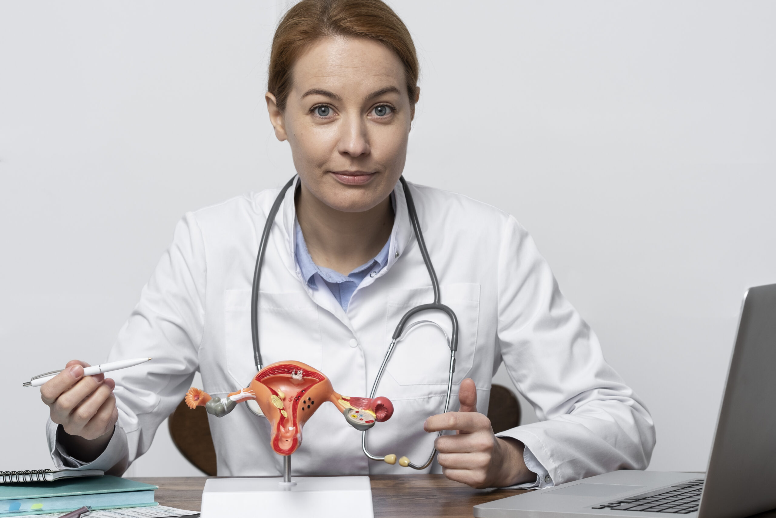
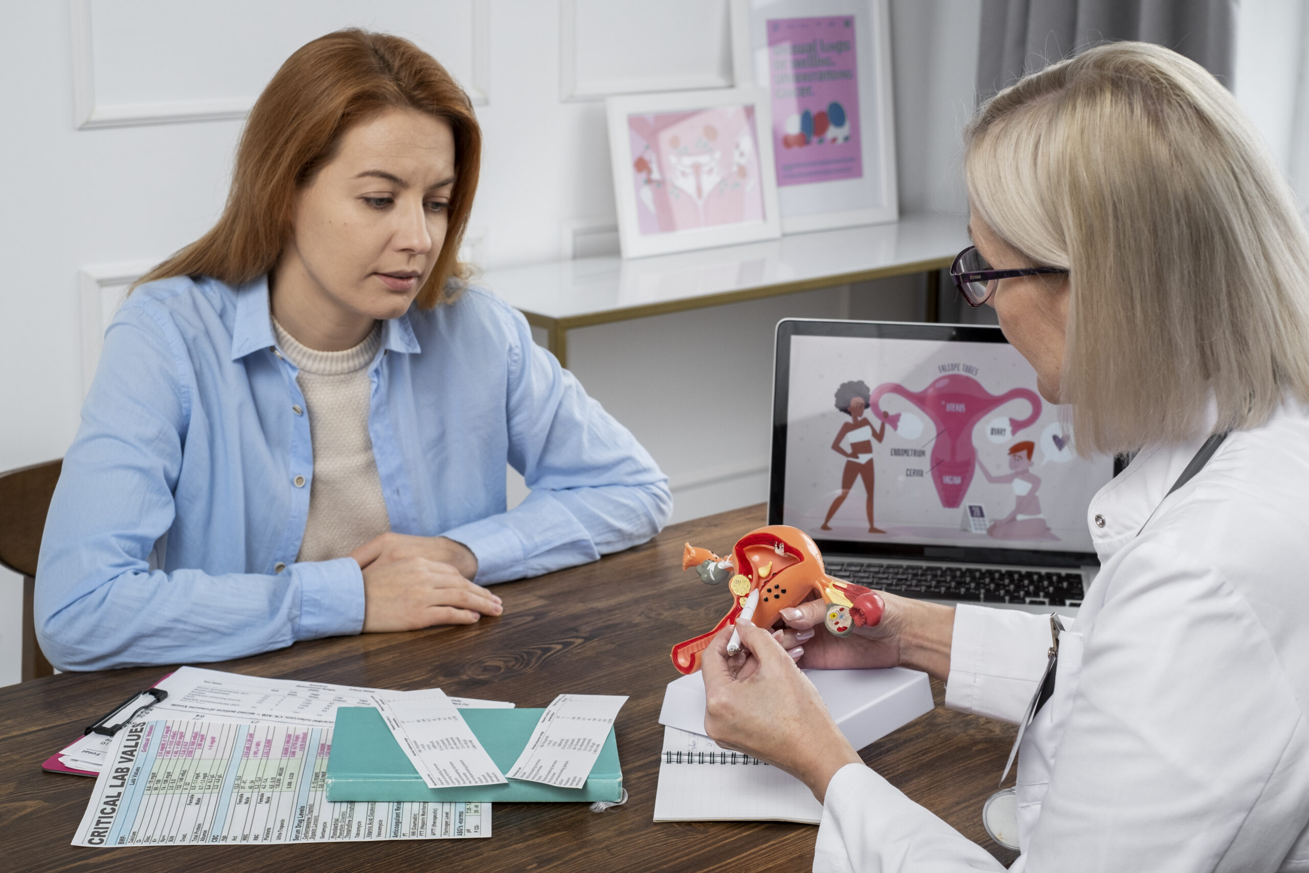
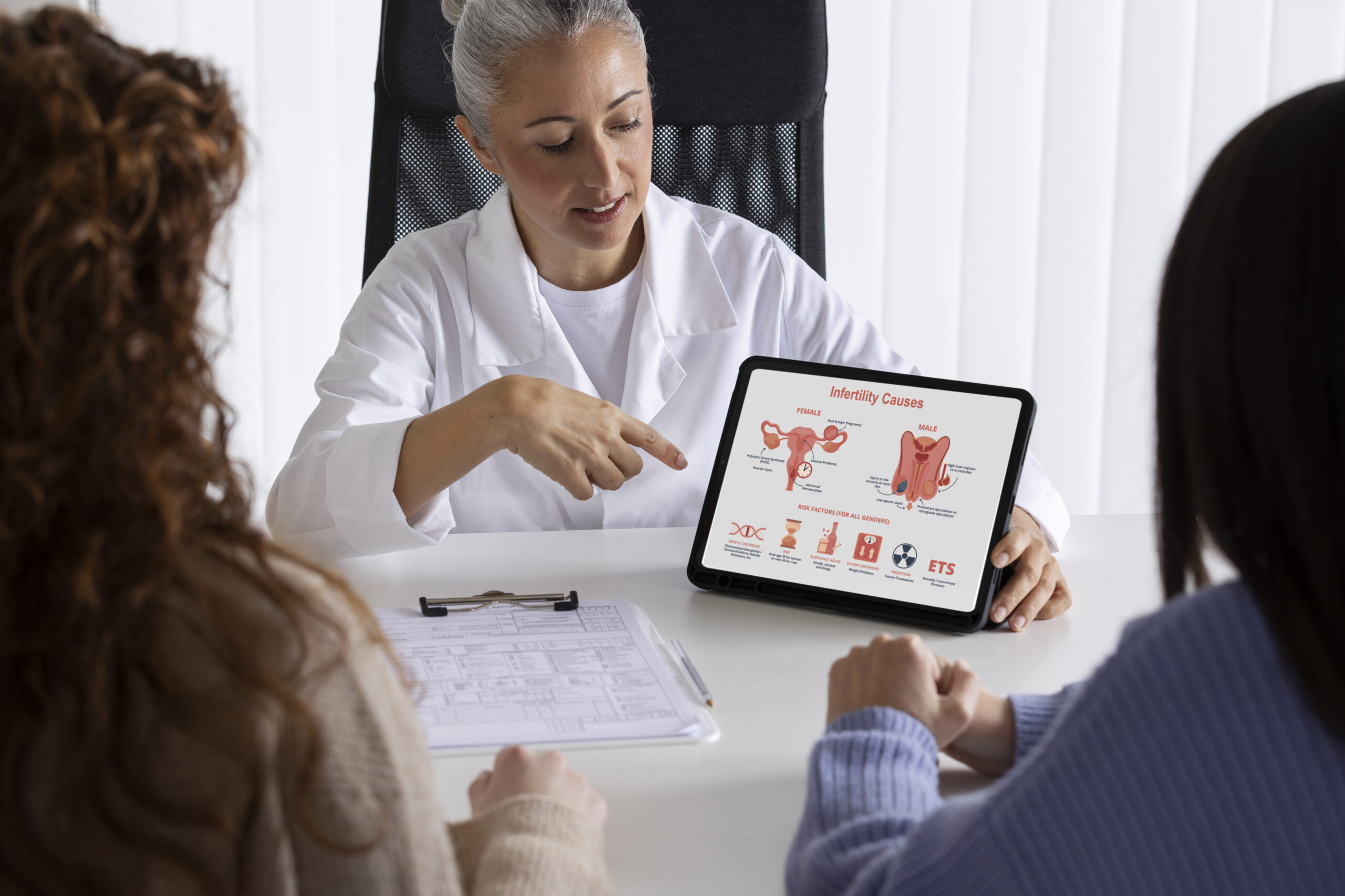
At Seamless Medical Centers, Dr. Zagum Bhatti, Board-Certified Interventional Radiologist, performs UFE and guides patients through recovery for patients across Southeast Texas and the Houston area. Both Houston-area UFE service information and Port Arthur UFE service information are available.
Immediately After the Procedure: The First 24 Hours
UFE is performed under conscious sedation, which means you will be comfortable during the procedure but will not require the extended recovery from general anesthesia. After the procedure, you will rest in a recovery area for several hours while your vital signs are monitored and the initial post-embolization response is assessed. Some patients go home the same day; others stay overnight for pain management.
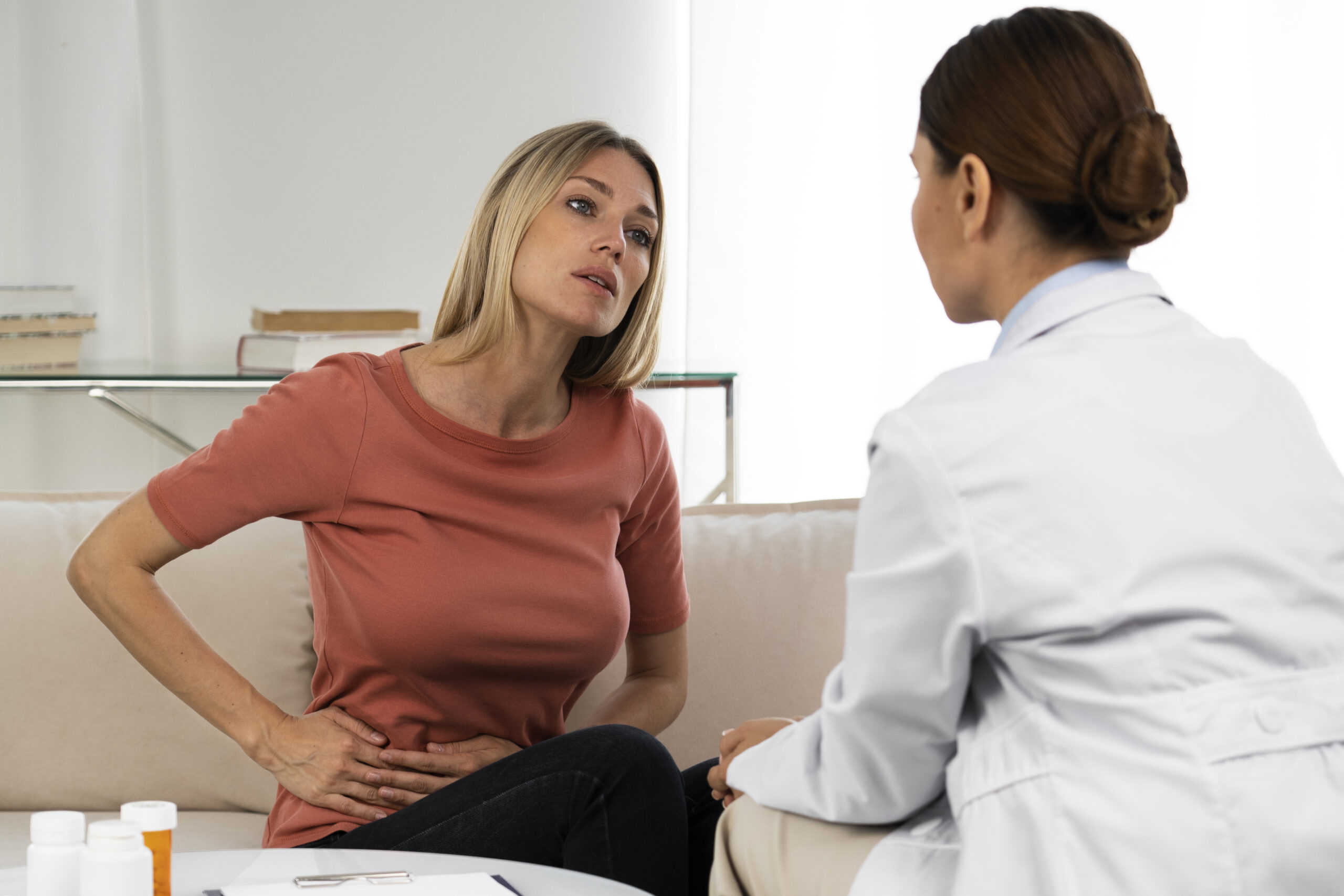
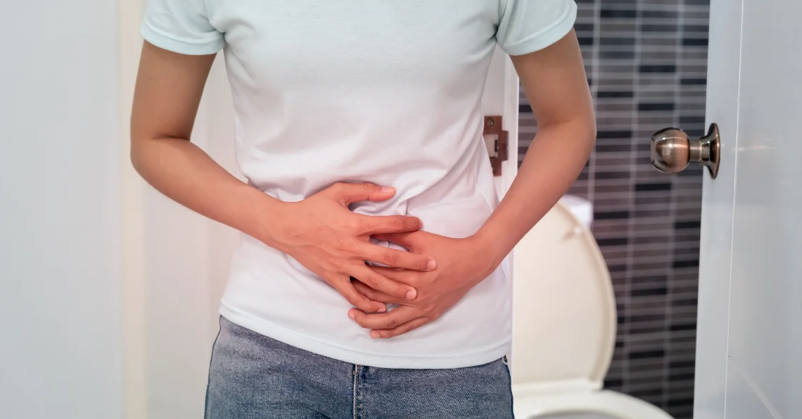
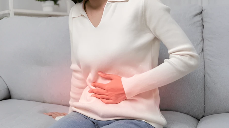
The most significant symptom in the first 24 hours is pelvic cramping — often described as intense menstrual cramping — that begins as the embolization takes effect and the fibroids are cut off from their blood supply. This cramping is managed with prescribed pain medications and typically peaks in the first 12 to 24 hours before beginning to decrease. You will need someone to drive you home and should plan to rest for the remainder of the day.
The access site in your wrist or groin will have a small dressing. Keep it clean and dry as directed. Minor bruising and tenderness at the site are normal and resolve over one to two weeks. You may also experience a low-grade fever, fatigue, and nausea in the first 24 to 48 hours as part of the post-embolization syndrome — a normal inflammatory response to the treated fibroids.
Days 2 Through 7: Post-Embolization Syndrome
The first week after UFE is characterized by what is called post-embolization syndrome: a cluster of symptoms including fatigue, mild fever (typically below 101°F), pelvic cramping or aching, vaginal discharge, and general malaise. These symptoms reflect the body’s response to the treated fibroids and are expected rather than alarming. They typically peak around day two to three and gradually improve through the first week.
Pain management during this phase usually transitions from stronger medications prescribed at discharge to over-the-counter anti-inflammatory medications as the cramping becomes more manageable. Most patients find they need strong pain medication for the first two to four days and manage adequately with ibuprofen or naproxen afterward. Staying ahead of pain with scheduled doses rather than waiting until pain is severe is generally more effective.
Activity during the first week should be light. Walking around your home, short walks outside, and light daily tasks are appropriate. Avoid strenuous exercise, heavy lifting, and prolonged sitting or standing. Most patients feel significantly better by the end of the first week and begin to feel more like themselves. If your fever exceeds 101°F, is accompanied by shaking chills, or if pelvic pain is worsening rather than improving after day three, contact the office.
Weeks 2 Through 4: Returning to Normal Activities
By week two, most patients have completed the acute recovery phase and are transitioning back to normal activities. Women with desk jobs or sedentary work often return to work during the second week. Women with physically demanding jobs typically need three to four weeks before returning to full duty.
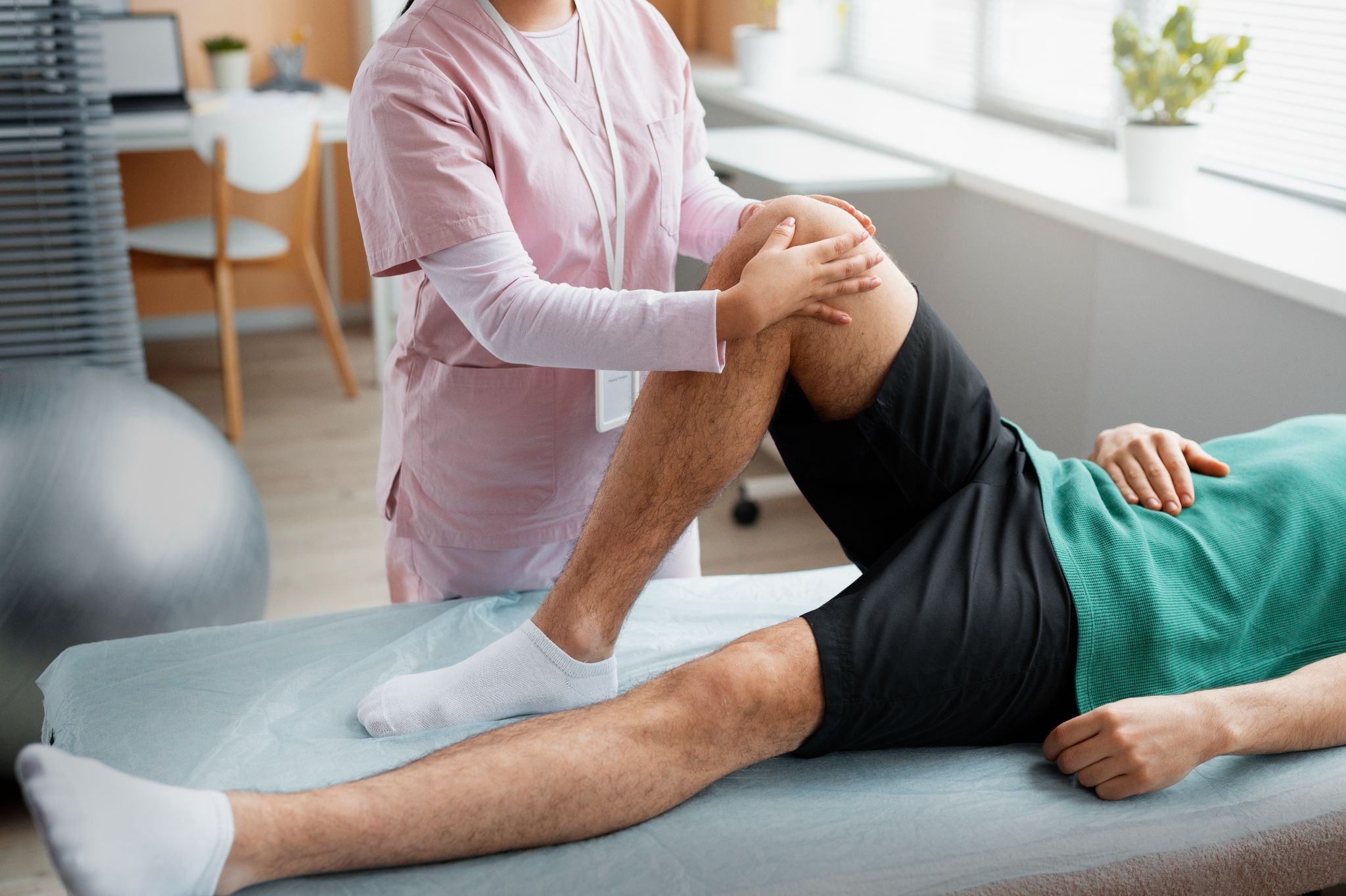
Exercise can resume gradually during weeks two to four. Walking is appropriate immediately. Light exercise such as yoga, stretching, and moderate cardio can typically resume by week two to three. More intense exercise, heavy lifting, and high-impact activities should wait until week four or until cleared by Dr. Bhatti at your follow-up appointment.
You may notice changes in your menstrual cycle beginning with your first period after UFE, which typically occurs four to six weeks after the procedure. Some women notice their first post-UFE period is heavier than usual as the uterine lining sheds residual fibroid tissue. Subsequent cycles should progressively become lighter and more regular as fibroids continue to shrink. If you have questions about what you’re experiencing, contact Seamless Medical Centers.
Months 2 Through 6: Maximum Benefit Develops
The full benefit of UFE develops over three to six months as fibroids continue to shrink. Most women experience significant improvement in heavy menstrual bleeding within the first one to two cycles after the procedure. Bulk symptoms — pelvic pressure, urinary frequency, abdominal fullness — improve as fibroid volume decreases. The extent of shrinkage depends on individual fibroid characteristics and vascular anatomy. Learn how UFE compares to other fibroid treatments to understand what makes UFE different from surgical options.
Follow-up imaging, typically a pelvic MRI or ultrasound, is usually performed three to six months after UFE to assess fibroid response. This imaging helps confirm the degree of shrinkage and identify any fibroids that may not have responded fully. Most women find that the combination of reduced bleeding, reduced bulk symptoms, and improved quality of life represents a meaningful improvement compared to their pre-treatment baseline.
When to Contact Seamless Medical Centers During Recovery
Contact the office promptly if you develop fever above 101°F that persists more than 48 hours after the procedure, if pelvic pain is worsening rather than improving after the first three days, if you notice foul-smelling vaginal discharge, if you develop increasing redness or swelling at the access site, or if you experience symptoms of urinary tract infection. For any emergency symptoms including severe pain unresponsive to medication, shortness of breath, or chest pain, call 911 or go to the emergency room.
Contact us with any questions during your recovery. Our team is available to help you determine whether what you’re experiencing is part of the normal recovery process or warrants evaluation.
Schedule Your Consultation
If you’re ready to explore your options, contact Seamless Medical Centers to schedule a consultation with Dr. Bhatti. Phone: 409-213-9575. Address: 3300 Jimmy Johnson Blvd, Suite #130, Port Arthur, Texas 77642.
Medical Disclaimer
Individual results may vary. This information is for educational purposes only and should not replace professional medical advice. Treatment decisions should be made in consultation with qualified healthcare providers.
Published by Seamless Medical Centers | Clinical information reflects the expertise of Dr. Zagum Bhatti, MD, Board-Certified Interventional Radiologist, Founder of Seamless Medical Centers.