Author: a_mustafa

  • Leg Pain: Types, Causes, Symptoms, Treatments & Prevention

    Leg Pain: Types, Causes, Symptoms, Treatments & Prevention

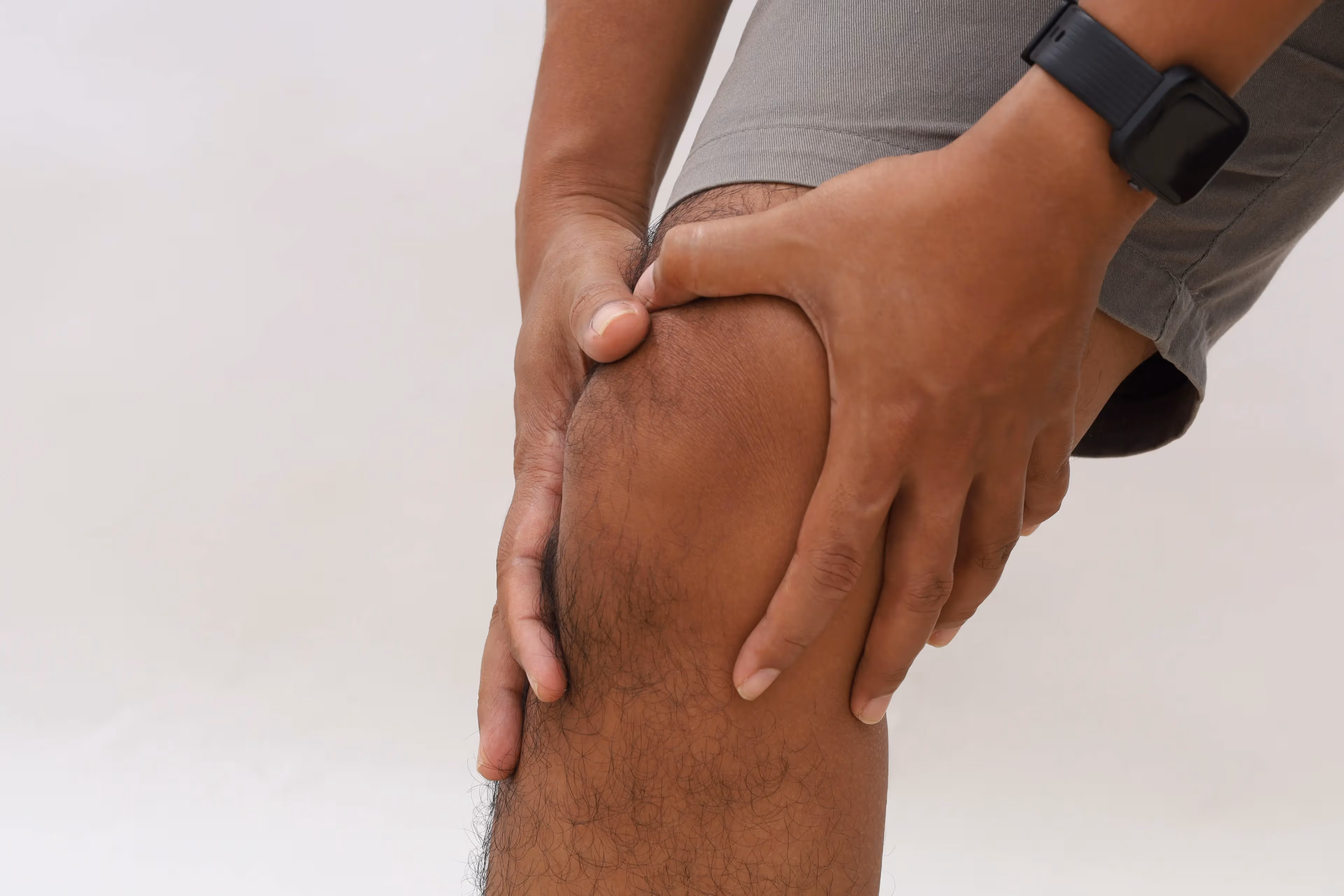
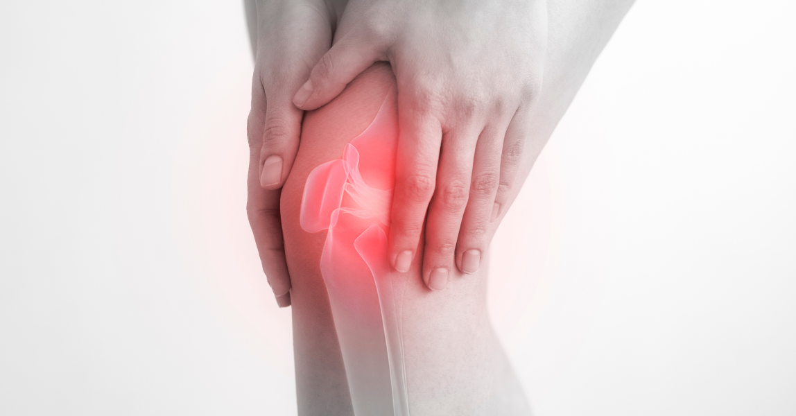
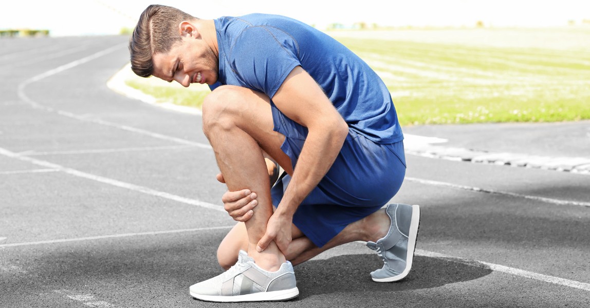
    Leg pain is a common yet often misunderstood symptom.

    While many people experience temporary aches after exercise, long work shifts, or standing all day, persistent or severe leg pain can be a sign of something more serious.

    In fact, leg pain is one of the most frequently misdiagnosed symptoms because it can stem from multiple structures, muscles, nerves, joints, bones, or blood vessels. This overlap often confuses.

    For example, peripheral artery disease (PAD) is commonly mistaken for back pain or sciatica, and some patients even undergo spine evaluations or treatments before the vascular cause is identified.

    Meanwhile, nerve-related pain, such as sciatica, can mimic vascular or muscular disorders.

    However, location, type of pain, and what triggers or relieves it are often the biggest clues: sharp, shooting pain often points toward nerve involvement; dull, aching pain suggests muscle or joint irritation; and cramping during walking may indicate circulation problems.

    To help patients, families, and caregivers, this guide clarifies what leg pain is, explores the major types of leg pain, reviews common causes, highlights red‑flag symptoms, and summarizes evidence‑based treatments and prevention strategies.
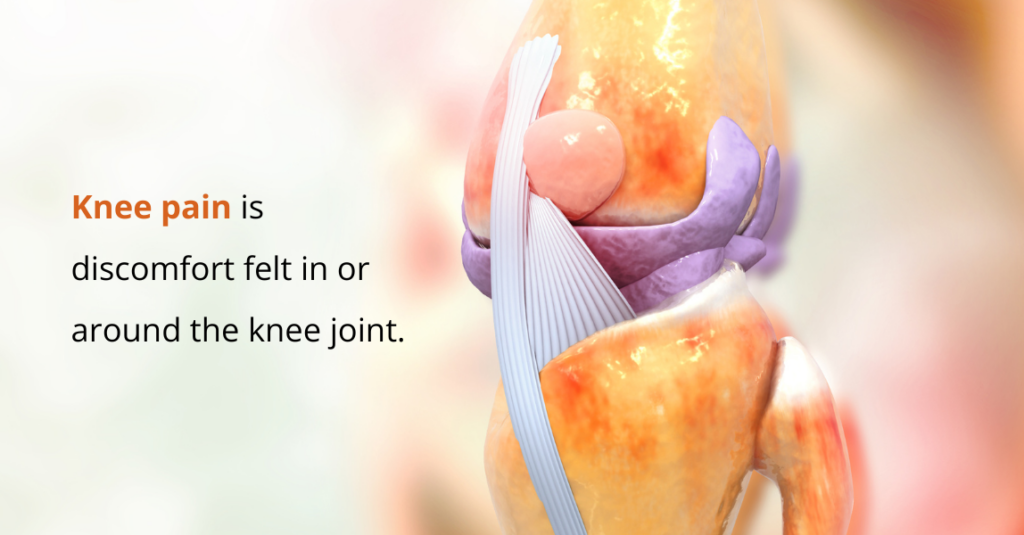
    What is Leg Pain?

    From a medical perspective, leg pain refers to any discomfort, soreness, or aching felt between the hip and the ankle. It can be acute (sudden and short-term) or chronic (lasting weeks or longer), and it may come and go or stay constant.

    People often describe leg pain as:

    • A sharp, shooting pain
    • A dull or heavy ache
    • A burning or tingling sensation
    • Cramping, especially at night.

    Leg pain can come from almost any structure in the leg, including your:

    • Bones
    • Muscles
    • Tendons and ligaments
    • Joints
    • Nerves
    • Blood vessels
    • Soft tissues

    Because so many tissues are involved, the cause of leg pain varies widely.

    However, it’s important to seek medical care if the pain is severe, persistent, or comes with swelling, numbness, colour changes, or difficulty walking.

    4 Types of Leg Pain

    types of leg pain

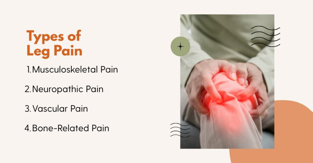
    Medical experts and researchers typically classify leg pain by the major tissue type or system affected, as this helps guide diagnosis and treatment.

    Here are the main types of leg pain:

    1. Musculoskeletal Pain (Muscle and Tendon)

    Musculoskeletal leg pain refers to discomfort that arises from muscles, tendons, ligaments, and other soft-tissue structures.

    They can occur due to:

    • Muscle strains: These injuries happen when a muscle is overstretched or torn. Strains are common, especially in athletes or during sudden increases in activity.
    • Shin splints: This condition involves irritation and inflammation of the muscles, tendons, or tissues surrounding the shin bone (tibia), often triggered by overuse.
    • Stress fractures: Tiny cracks in the bone caused by repetitive force or impact, often seen in runners and other high-impact sports.
    • Tendinitis: Also called tendonitis, this occurs when the tendon connecting muscle to bone becomes inflamed, typically from overuse or repetitive motion.

    2. Neuropathic Pain (Nerve-Related)

    Neuropathic leg pain occurs when the nerves are irritated, damaged, or compressed. Conditions such as sciatica, nerve compression, and neuropathy commonly trigger this type of pain.

    Unlike muscle-related discomfort, neuropathic pain follows nerve pathways and often radiates from the lower back or hip into the leg.

    People typically describe neuropathic pain with distinct sensations, including:

    • Burning pain
    • Tingling or pins and needles
    • Shooting or electric-shock sensations
    • Numbness
    • Sometimes, leg weakness or reduced sensation

    These features help differentiate nerve-related pain from musculoskeletal pain and often indicate an underlying neurological issue that requires proper evaluation.

    A prospective study of patients with low-back–related leg pain found that depending on the definition used, 48% to 74% of these cases showed neuropathic pain features.

    3. Vascular Pain (Blood-Flow/Circulation-Related Pain)

    Vascular leg pain occurs when blood flow to or from the leg is impaired.

    Typical features of vascular pain include:

    • Varicose veins: These develop when blood pools inside the veins, causing them to enlarge and appear blue or purple beneath the skin. They may lead to heaviness, aching, or swelling in the legs.
    • Peripheral artery disease (PAD): PAD occurs when plaque made of fat and cholesterol builds up in the arteries, narrowing them and reducing blood supply to the legs. This often causes cramping or aching during activity.
    • Deep vein thrombosis (DVT): DVT is a blood clot in a deep vein that blocks normal blood flow. It can cause swelling, warmth, and tenderness in one leg. This condition is serious and requires prompt medical attention.

    A recent biomechanical study showed that people with PAD have impaired gait, even when not currently in pain, which underlines how vascular problems affect leg function beyond just subjective discomfort.

    4. Bone-Related Pain

    Bone-related leg pain originates from problems in the bone itself, such as stress fractures, shin splints, or arthritis.

    The underlying mechanism is usually: repetitive mechanical stress → micro-damage to bone → bone irritation or fractures → ongoing pain.

    This type of pain is typically deep, persistent, and often worsens with impact or weight-bearing activities like walking or running. It can also occur due to:

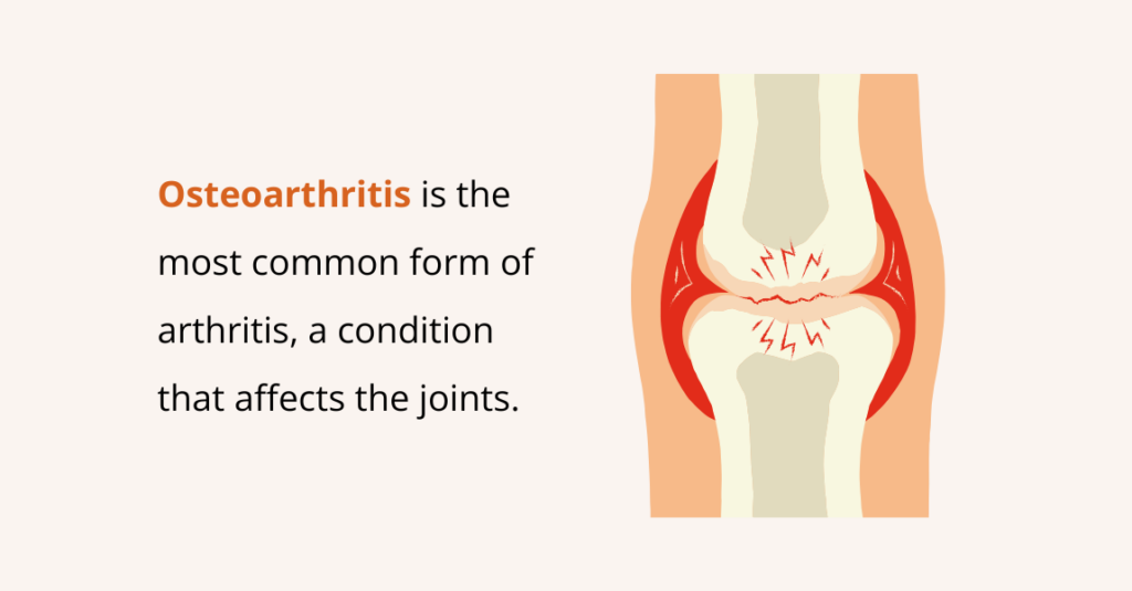
    • Arthritis: A very common condition characterized by joint pain, stiffness, and inflammation. There are more than 100 different types, affecting people of all ages.
    • Gout: A form of inflammatory arthritis that causes sudden episodes of intense joint pain and swelling. It occurs when uric acid builds up in the body and forms sharp crystals in the joints.

    Although many reviews separate bone and joint pain from soft-tissue musculoskeletal pain, classic orthopedic and sports-medicine literature lists stress fractures and bone-stress injuries as frequent causes of chronic leg pain, especially in athletes or people who are increasing their activity.

    Because these injuries can worsen if left untreated, proper diagnosis, often including imaging, and load management, such as rest or reduced-impact activity, are essential to prevent further damage.

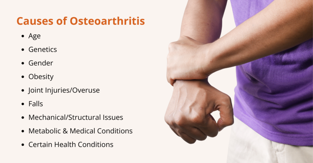
    What Causes Leg Pain?

    what causes of leg pain

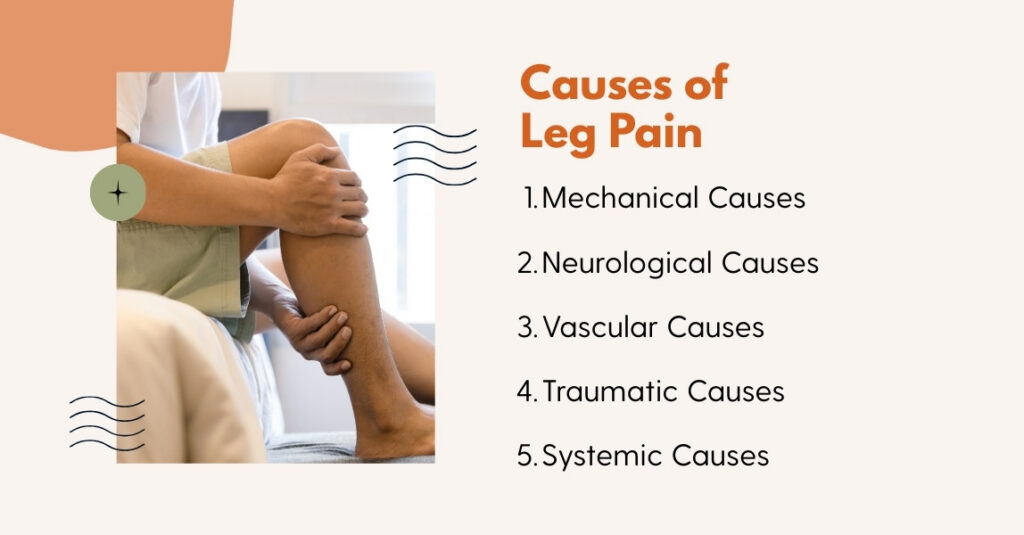
    Leg pain can arise from a wide range of mechanical, neurological, vascular, traumatic, and systemic factors. Thus, understanding the underlying cause is essential for accurate treatment and prevention.

    1. Mechanical Causes

    Mechanical issues occur when the muscles, bones, or joints are stressed beyond their capacity.

    Overuse injuries and muscle imbalances are common: repetitive activity, sudden increases in training, or weak supporting muscles can lead to strains, tendinitis, or stress fractures.

    Even sedentary jobs can lead to stiffness and cramps when activity resumes.

    Improper footwear and flat feet also contribute. Also, poor arch support alters biomechanics, placing extra stress on muscles and joints and increasing the risk of shin splints or plantar fasciitis.

    2. Neurological Causes

    Leg pain can stem from nerve problems. Herniated discs or spinal stenosis compress spinal nerves, leading to sciatica, burning, tingling, or numbness in the leg.

    Nerve entrapment conditions, such as peroneal neuropathy, may cause weakness or difficulty lifting the foot.

    Moreover, prolonged sitting or poor posture can tighten hip flexors, compressing nerves and contributing to neuropathic leg pain.

    3. Vascular Causes

    Circulatory problems are another major source of leg pain. Peripheral Artery Disease (PAD) occurs when atherosclerosis narrows leg arteries, causing cramping or aching during walking or activity.

    The risk factors include smoking, diabetes, hypertension, and high cholesterol. The symptoms include unilateral swelling, tenderness, warmth, or a “pulling” sensation in the calf.

    Research shows that varicose veins and venous insufficiency result from prolonged standing or genetic factors, leading to leg heaviness, cramps, and visible veins. Roughly one in four adults may have visible varicose veins.

    4. Traumatic Causes

    Direct trauma from falls, accidents, or sports injuries can cause fractures, sprains, or contusions.

    If you experience this, seek immediate care if you hear a popping sound, notice a visible deformity, or are unable to bear weight.

    Similarly, compartment syndrome is a serious condition where swelling within muscle compartments causes severe pain and requires emergency surgery to prevent permanent damage.

    5. Systemic or Metabolic Causes

    Diabetes and neuropathy can damage nerves and blood vessels, producing burning, tingling, and numbness in the legs. Also, inflammatory joint conditions like arthritis or gout can cause deep joint pain, stiffness, and swelling.

    Moreover, infections, including cellulitis or osteomyelitis (bone infection), may produce redness, warmth, and pain. Fever accompanied by other symptoms is a red flag that requires urgent medical attention.

    Kristofer Jones, MD, an orthopaedic surgeon at UCLA Health, says that many people with sedentary jobs cram all their physical activity into the weekend. This sudden spike in load can cause strains or stress fractures; he recommends gradually increasing activity, varying exercises, and building core strength. Ignoring early discomfort can turn a minor strain into a serious injury.

    The “Weekend-Warrior-Mentality”

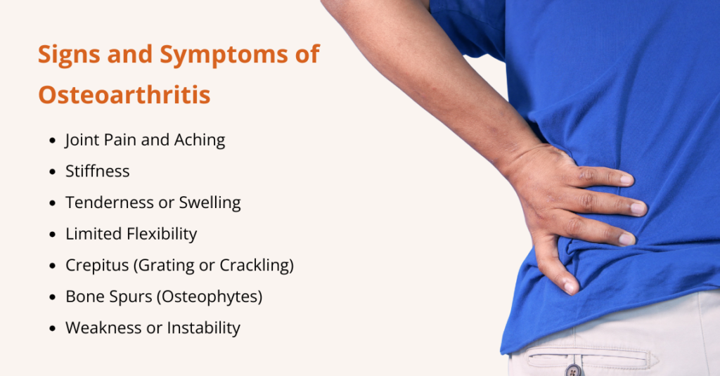
    What are the Symptoms of Leg Pain?

    Leg pain can appear in many forms. It may affect a small, specific area or spread across the entire leg. The discomfort can also extend into the buttocks, foot, lower back, or spine, depending on the cause.

    In some cases, pain occurs in multiple body regions at the same time. The pain may feel:

    • Dull or sharp
    • Burning or tingling
    • Your leg may also feel numb

    It can be triggered by activity (for example, walking or running), by position (standing or sitting), or by coming on at night. Common features associated with leg pain include swelling, numbness, colour changes, warmth or redness, and difficulty walking.

    Additional signs and symptoms that can accompany leg pain include:

    • Swelling, especially if one leg looks noticeably different from the other
    • Varicose veins
    • Sores or ulcers
    • Redness/warmth
    • Numbness/sensory change
    • Color changes in the leg or foot
    • Slow-healing wounds
    • General unwellness, especially during recovery from an infection or fracture

    It is important to seek medical attention if your leg pain is sudden, severe, persistent, or if you experience any of these additional symptoms.

    Vascular surgeon George Anton, MD, notes that visible venous disease is not only cosmetic, “When veins are big enough, the blood that pools in them can also clot. Clots can travel through your body, putting you at risk for a pulmonary embolism, which could be life-threatening.”

    Clinical Insight

    Leg Pain Treatments

    leg pain treatment options

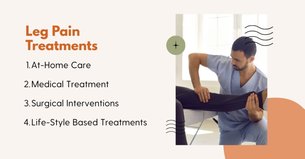
    Leg pain treatment depends on the cause, but many mild issues improve with home care, such as gentle stretching, movement, and simple pain relievers like paracetamol or ibuprofen.

    More serious causes may require antibiotics, targeted medication, or, in some cases, surgery.
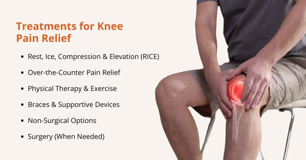
    At-Home Care

    For many mild leg problems, especially muscle, tendon, or soft-tissue pain, at-home care can help relieve pain and support healing.

    Some of the common approaches include:

    • RICE (Rest, Ice, Compression, Elevation)

    Rest the leg, apply ice, use gentle compression (e.g., bandage), and elevate the leg above heart level. Research shows that RICE can reduce pain and swelling after a strain, sprain, or minor injury.

    • Stretching and hydration

    Gentle stretching helps keep muscles and tendons flexible. Staying well-hydrated supports muscle health and may reduce the chance of cramps or overuse injury.

    • Epsom salt soaks

    Warm salt baths (when no open wounds) can help ease muscle soreness and promote relaxation, useful for muscle or tendon discomfort.

    According to a study, Epsom salt has anti-inflammatory and analgesic properties, making it a pain-relieving agent.

    • Over-the-counter (OTC) pain relievers/anti-inflammatories

    Drugs like non-steroidal anti-inflammatory drugs (NSAIDs) or acetaminophen help relieve pain, reduce inflammation, and make movement easier.

    These “home-care” methods often work for sprains, strains, mild tendon irritation, or post-activity soreness. If pain persists or worsens, seeking medical care is advised.

    Medical Treatments

    When at-home care isn’t enough, or when leg pain stems from vascular, nerve, or deeper structural problems, medical treatments may be required:

    • Physical therapy

    A physical therapist can guide gentle movement, strengthen muscles, improve flexibility, and correct movement patterns. This helps many people with musculoskeletal or nerve-related leg pain recover without surgery.

    • Anti-inflammatory medications(NSAIDs)

    For pain and inflammation, NSAIDs remain a first-line treatment. They reduce swelling and relieve discomfort in soft-tissue injuries, arthritis, or after surgery.

    • Antibiotics

    If leg pain stems from an infection (e.g., cellulitis, infected joint, bone infection), antibiotics are required. Infections accompanied by redness, warmth, fever, or spreading pain warrant prompt medical treatment.

    • Nerve blocks or nerve-directed treatments

    For severe nerve-related pain (nerve compression, radiculopathy), nerve-block injections or other nerve-targeted therapies may be recommended, especially when conservative measures fail.

    • Blood thinners (anticoagulants)

    If leg pain is due to a blood clot (deep vein thrombosis, DVT), anticoagulant (“blood thinner”) medications are standard treatment to prevent clot growth or migration.

    For vascular problems like DVT or chronic venous insufficiency, compression therapy (compression stockings or wraps) may also be prescribed to improve blood flow and reduce swelling.

    Surgical or Procedural Interventions

    When conservative or medical treatments are insufficient, surgery or interventional procedures may become necessary, for example:

    • Fractures, severe bone damage, or structural joint problems may require surgical repair or stabilization.
    • Severe nerve compression (e.g., spinal nerve-root compression) may require surgical decompression.
    • Vascular blockages, clots, or severe venous/arterial disease may require vascular intervention, such as clot removal, angioplasty, or vein surgery (depending on the condition).

    In post-surgical situations, pain medications and physical therapy often support recovery. It is evident that NSAIDs, acetaminophen, and pain-management strategies help reduce pain after surgery.

    Lifestyle-Based & Preventive Treatments

    Prevention and long-term management often depend on lifestyle and biomechanical adjustments:

    • Custom orthotics

    For foot or gait-related leg pain (e.g., flat feet, shin splints, tendon issues), custom orthotics or insoles can correct biomechanics, reduce abnormal stress, and prevent recurrence.

    • Strength building and regular exercise

    Strengthening muscles, improving flexibility, and balancing muscle groups help support joints and bones, reducing the risk of overuse injuries, tension, or biomechanical stress that can lead to pain.

    • Weight management

    Maintaining a healthy weight reduces load on bones, joints, and vessels, lowering the risk of arthritis, vascular disease, or overuse injuries.

    • Smoking cessation

    For vascular-related leg pain (like peripheral artery disease, PAD), stopping smoking is critical to improve blood flow and slow disease progression.

    Also, for those at risk of vascular problems or blood clots, lifestyle habits, such as regular movement, avoiding prolonged immobility (e.g., on long flights), maintaining a healthy diet, and staying hydrated, help reduce risk.

    How to Prevent Leg Pain?

    You can lower your risk of leg pain by stretching before and after physical activity, staying hydrated, and eating potassium-rich foods like bananas and chicken to support healthy muscles and tendons.

    To prevent health conditions that can cause leg nerve damage:

    • Maintain overall health: Get enough sleep, stay hydrated, and eat a balanced diet rich in potassium, magnesium, and calcium to support muscle and nerve function.
    • Exercise regularly and gradually: Include aerobic, strength, and flexibility training, increasing intensity slowly to avoid overuse injuries.
    • Warm-up and cool-down: Gentle stretching before and after activity helps prevent strains.
    • Wear proper footwear: Good arch support and cushioning reduce stress on joints and bones.
    • Maintain a healthy weight and avoid smoking: Both reduce strain on the circulatory system and lower the risk of PAD and varicose veins.
    • Monitor key health markers: Keep blood pressure, cholesterol, and blood sugar within healthy ranges through regular check-ups.
    • Take movement breaks: Avoid prolonged sitting or standing to improve circulation and reduce nerve compression.
    • Listen to your body: Stop activity if you notice unusual pain, swelling, or numbness and seek timely medical care.

    Frequently Asked Questions (FAQs)

    When should someone see a doctor?

    You should seek medical attention if you experience any of the following:

    • Pain lasting more than 3–5 days without improvement
    • Severe or worsening pain that interferes with daily activities
    • Swelling in one or both legs, especially if sudden or accompanied by redness or warmth
    • Numbness, tingling, or weakness in the leg
    • Pain after an injury such as a fall, twist, or direct impact
    • Red flags like inability to bear weight, fever, or sudden color changes in the leg

    Prompt evaluation is important to identify underlying issues such as nerve compression, blood clots, vascular problems, or serious musculoskeletal injuries.

    Is Leg Pain Always from Injury?

    No, leg pain is not always caused by an injury. While strains, sprains, or fractures are common sources of leg pain, it can also arise from nerve, spinal, or vascular issues.

    • Nerve-related causes: Conditions such as sciatica, peripheral neuropathy, or spinal nerve compression can cause burning, tingling, shooting pain, or numbness in the leg, even without trauma.
    • Spinal issues: Herniated discs or spinal stenosis can compress nerves, causing pain to radiate down the leg. Often, patients feel leg pain more than back pain.
    • Vascular causes: Problems with blood flow, including peripheral artery disease (PAD), deep vein thrombosis (DVT), or varicose veins, can lead to aching, heaviness, swelling, or color changes. These pains may worsen with activity or prolonged standing and are unrelated to muscle injury.

    Understanding the underlying cause is crucial because treatment differs depending on whether the pain comes from muscles, nerves, bones, or blood vessels.

    Why Does Leg Pain Worsen at Night?

    Leg pain often feels worse at night due to several factors:

    • Circulation changes: When you lie down, blood flow and venous return in the legs can shift, sometimes worsening discomfort from conditions like peripheral artery disease (PAD) or varicose veins.
    • Nerve irritation: Nerve-related pain, such as sciatica or peripheral neuropathy, can become more noticeable at rest because there are fewer distractions and the body is more aware of discomfort.
    • Restless Leg Syndrome (RLS): This neurological condition causes an uncontrollable urge to move the legs, often accompanied by aching, tingling, or crawling sensations, which typically intensify in the evening or at night.

    Recognizing the pattern and timing of leg pain can help clinicians identify the underlying cause and guide appropriate treatment.

    Conclusion

    Leg pain is a symptom with many faces. While minor aches after exercise are common, persistent or severe pain may indicate underlying musculoskeletal, neurologic, vascular, or systemic disorders.

    Early recognition of symptoms and prompt medical evaluation are crucial, especially for red flags like sudden swelling, color changes, or neurological deficits.

    Evidence‑based treatments range from at‑home RICE therapy to physical therapy, medications, surgery, or anticoagulation, depending on the cause.

    Moreover, preventive strategies such as regular exercise, proper footwear, smoking cessation, and careful attention to cardiovascular health can substantially reduce the risk of leg pain.

    Remember that an accurate diagnosis is the key to effective treatment.

    Consult healthcare professionals rather than self‑diagnosing, and take leg pain seriously; it might be your body’s warning of a more significant health issue.

  • Knee Osteoarthritis: Causes, Symptoms, Diagnosis, Treatment, & Prevention

    Knee Osteoarthritis: Causes, Symptoms, Diagnosis, Treatment, & Prevention

    Knee osteoarthritis (OA) is a common and growing public health concern in the United States.

    As people live longer and rates of obesity rise, more Americans are developing this degenerative joint disease, leading to major personal and societal costs.

    In fact, research estimates that about 14 million U.S. adults suffer from symptomatic knee OA.

    Moreover, radiographic studies from the U.S. show that more than one in three adults aged 60 or older have signs of knee OA on imaging, and around 12 percent report symptoms.

    As the population ages and obesity continues to climb, resulting in knee OA prevalence rising further, understanding risk factors, diagnosis, and treatment is more important than ever.

    In this article, we’ll walk through what knee osteoarthritis is, how it progresses, who’s most at risk, what causes it, how it’s diagnosed, treatment options, and finally, strategies for prevention.

    What is Knee Osteoarthritis?

    what is knee osteoarthritis

    Knee osteoarthritis occurs when the cartilage that cushions your knee joint gradually wears down. As this protective layer thins, the bones begin to rub against each other, creating friction that leads to pain, swelling, and stiffness.

    Because the knee carries your body weight and absorbs impact with every step, it’s one of the joints most commonly affected by this degenerative “wear-and-tear” disease.

    Research shows that OA of the knee joint is often associated with aging, but it can also be influenced by factors such as injury, obesity, genetics, and other health conditions.

    As the condition worsens, individuals may experience difficulty performing daily activities, such as walking, climbing stairs, or getting up from a chair. Over time, though, the disease can worsen and may eventually limit mobility.

    However, many treatments can help slow its progression, reduce pain, and improve everyday function. Healthcare providers carefully monitor its progression, and if knee OA begins to significantly affect your quality of life, surgical and non-surgical options are available.

    What are the Stages of Knee Osteoarthritis?

    what are the stages of knee osteoarthritis

    Although there’s no single universal staging system, knee osteoarthritis typically follows a predictable, gradual course.

    Clinicians often describe it in stages to explain how advanced the disease is and what to expect.

    Knowing these stages can help you recognize symptoms early and plan treatment with your healthcare team.

    • Stage 1 (Minor)

    At this earliest stage, there is minor wear and tear to the cartilage. Changes are usually subtle, and most people feel little or no pain. X-rays may show only very subtle changes, if any at all.

    • Stage 2 (Mild)

    Mild OA means the cartilage is beginning to break down. You may notice occasional pain or stiffness, especially after activity or when getting up from a seated position. There is still enough cartilage to prevent bones from rubbing together, but symptoms are starting to appear.

    • Stage 3 (Moderate)

    In the moderate stage, cartilage loss is more pronounced. Pain becomes more frequent and may interfere with daily activities such as walking, climbing stairs, squatting, or kneeling. Stiffness and reduced range of motion are common, and you may feel discomfort after periods of rest.

    • Stage 4 (Severe)

    Severe OA indicates that cartilage is nearly or completely gone in parts of the knee. Bones may grind together, causing constant pain, swelling, marked stiffness, and significant limitations in movement. At this point, conservative measures often provide limited relief, and joint replacement surgery may be discussed as an option.

    What Causes Knee Osteoarthritis?

    what are the causes of knee osteoarthritis

    Knee osteoarthritis is caused by the gradual breakdown of cartilage in the knee joint.

    While the exact cause of this breakdown is not always clear, several factors contribute to its development:

    • Mechanical Stress: Research shows that overuse of the knee joint, especially among individuals who engage in repetitive activities or heavy lifting, can cause cartilage wear and tear.
    • Inflammation: Chronic joint inflammation, often due to conditions such as rheumatoid arthritis, can accelerate cartilage breakdown.
    • Genetics: Modern research shows genetic factors can contribute to the development of knee osteoarthritis. If you have a family history of the condition, you may be more likely to develop it yourself.
    • Joint Instability: Knee instability due to prior injuries or muscle strength imbalances can place additional stress on the joint, leading to cartilage damage.
    • Obesity: Studies show that excess body weight puts additional pressure on the knee joint, leading to increased cartilage wear and tear.

    What are the Symptoms of Knee Osteoarthritis?

    The symptoms of knee osteoarthritis can vary depending on the severity of the condition.

    Knee pain is the most common and noticeable symptom of knee osteoarthritis. You may feel discomfort when putting weight on your knee, during movement, or even while resting.

    As the condition progresses, other symptoms can appear, including:

    • Stiffness, especially when you first wake up or after sitting for long periods.
    • Swelling or a puffy feeling around the knee joint.
    • Cracking, popping, or grinding sounds (called crepitus) when you move the knee.
    • A feeling of instability, as if the knee might buckle or “give out.”
    • Locking or catching, where the knee briefly feels stuck or difficult to move.

    These symptoms may come and go at first, but often become more frequent over time as osteoarthritis progresses.

    Who is at Risk for Knee Osteoarthritis?

    Several factors can increase the likelihood of developing knee osteoarthritis. Some of the key risk factors include:

    • Age: As we age, the cartilage in our joints naturally wears down, increasing the risk of developing OA. It is most common in people over 50.
    • Gender: Women are more likely than men to develop knee osteoarthritis, especially after menopause. This may be due to hormonal changes that affect the joints.
    • Obesity: Excess body weight adds stress to the knee joint, increasing the risk of cartilage wear. Studies show that individuals with a body mass index (BMI) of 30 or higher are at significantly higher risk of developing knee OA.
    • Previous Joint Injury: Individuals who have experienced a knee injury, such as a ligament tear or fracture, are at higher risk for developing OA later in life. Even if the injury heals, the knee joint may be weakened, leading to cartilage damage.
    • Genetics: Some people may have a genetic predisposition to osteoarthritis. Certain genes may make individuals more susceptible to cartilage breakdown in the knee joint.
    • Occupation: Jobs that require heavy lifting, kneeling, or squatting can increase the risk of developing knee osteoarthritis due to repetitive stress on the joint.
    • Other Health Conditions: Conditions such as rheumatoid arthritis, gout, and diabetes can also increase the likelihood of developing knee osteoarthritis.

    How is Knee Osteoarthritis Diagnosed?

    how is knee osteoarthritis diagnosed

    Knee osteoarthritis is typically diagnosed through a combination of physical examination, medical history, and imaging.

    The following steps may be involved:

    • Physical Examination: Your doctor will assess your knee’s range of motion, check for swelling or tenderness, and evaluate how well your knee functions during movement.
    • Medical History: Your doctor will ask about your symptoms, any previous knee injuries, and your family history of osteoarthritis or other joint disorders.
    • X-rays: X-rays are commonly used to evaluate the extent of joint damage and cartilage loss. They can show narrowing of the joint space, bone spurs, and other signs of OA.
    • MRI: An MRI may be used if your doctor needs more detailed images of the soft tissues in your knee, including cartilage, ligaments, and tendons.
    • Blood Tests: In some cases, blood tests may be done to rule out other conditions, such as rheumatoid arthritis, that may cause similar symptoms.

    Knee Osteoarthritis Treatment Options

    Knee osteoarthritis treatment focuses on relieving pain, improving mobility, and slowing the progression of joint damage.

    Options range from lifestyle changes and nonsurgical therapies to surgical procedures. In most cases, healthcare providers begin with conservative (nonsurgical) treatments before considering surgery.

    Nonsurgical treatments for knee OA

    Nonsurgical options aim to reduce pain and inflammation, support joint function, and delay the need for surgery. Common treatments include:

    • Pain Medications: Over-the-counter NSAIDs (such as ibuprofen) or acetaminophen can help reduce pain and swelling. Prescription options may be used if symptoms are more severe.
    • Physical therapy: Targeted exercises strengthen the muscles around the knee, improve joint stability, and increase flexibility, making daily activities easier.
    • Weight loss (when needed): Reducing body weight decreases stress on the knee joint. Even losing 5–10% of body weight can significantly reduce symptoms.
    • Knee braces: Braces help support the joint, reduce strain, and may shift pressure away from the most damaged part of the knee.
    • Corticosteroid (steroid) injections: These injections reduce inflammation and can provide short-term pain relief, especially during flare-ups.
    • Knee gel injections (viscosupplementation): Hyaluronic acid “gel shots” add lubrication inside the joint to improve movement and reduce pain.
    • Genicular nerve block injections: These target the small nerves around the knee to interrupt pain signals temporarily.
    • Platelet-rich plasma (PRP) injections: PRP uses concentrated platelets from your own blood to promote healing and reduce joint inflammation.
    • Genicular artery embolization (GAE): A minimally invasive procedure that reduces blood flow to inflamed tissues around the knee, helping decrease pain and swelling for some patients.

    Surgical treatments for knee OA

    Surgery is typically considered when pain is severe, daily function is limited, or nonsurgical treatments are no longer effective. Surgical options include:

    • Cartilage restoration or replacement: Techniques such as microfracture, grafting, or implanting new cartilage aim to repair small areas of damage in younger, active patients.
    • Knee bone reshaping (osteotomy): Surgeons cut and realign the bones around the knee to shift weight away from the damaged area, helping relieve pain and delay joint replacement.
    • Partial knee replacement: Only the damaged portion of the knee is replaced with an artificial implant. This option preserves more of your natural knee and may lead to a faster recovery.
    • Total knee replacement: The entire knee joint is replaced with artificial components. This is the most common surgical treatment for advanced knee OA and can provide long-lasting pain relief and improved mobility.

    How Can Knee Osteoarthritis Be Prevented?

    While you cannot entirely prevent knee osteoarthritis, there are several steps you can take to reduce your risk and protect your knee joint:

    • Exercise Regularly: Engaging in low-impact activities like swimming, cycling, and walking can help strengthen the muscles around the knee and improve joint stability.
    • Maintain a Healthy Weight: Reducing excess weight will reduce the pressure on your knees and help protect against OA.
    • Avoid Joint Injuries: Protect your knees during physical activities, especially high-impact sports, by using proper techniques and wearing protective gear.
    • Strengthen Your Muscles: Strengthening the muscles around the knee joint can help support the joint and reduce the risk of developing osteoarthritis.
    • Stretch Regularly: Regular stretching can improve flexibility and reduce stiffness in the knee joint.

    With these prevention strategies and staying proactive about your knee health, you can reduce your risk of developing knee osteoarthritis and maintain a high quality of life for years to come.

    Frequently Asked Questions (FAQs)

    What does knee osteoarthritis feel like?

    Knee osteoarthritis often feels like a combination of pain, stiffness, and joint grinding. You may notice aching pain during movement or after activity, stiffness when you first wake up or stand after sitting, and a cracking or grating sensation (crepitus) when the knee moves. Many people also experience knee swelling, a feeling that the knee is unstable or might “give out,” and occasional locking or catching when the joint gets stuck. As the condition progresses, you may find it harder to fully bend or straighten your knee, making everyday activities more difficult.

  • Benign Prostatic Hyperplasia: Causes, Symptoms, & Treatments

    Benign Prostatic Hyperplasia: Causes, Symptoms, & Treatments

    Benign Prostatic Hyperplasia (BPH) is the most common prostate condition affecting men, particularly as they age.

    In the United States, over 50% of men over 50 experience symptoms of BPH, and this number rises to 90% by age 85.

    BPH is closely linked with male lower urinary tract symptoms (LUTS), but it does not raise the risk of prostate cancer or cause sexual dysfunction. However, it can affect quality of life by leading to bothersome and sometimes embarrassing urinary issues.

    While age and genetics play a key role in the development of BPH, emerging research shows that modifiable factors, including hormone levels, obesity, glucose regulation, diet, exercise, and inflammation, also influence its onset and progression.

    Therefore, understanding these risk factors, recognizing early symptoms, and seeking timely diagnosis and treatment can dramatically improve outcomes and quality of life.

    This article provides an overview of BPH, including its causes, symptoms, risk factors, diagnosis, and the latest treatment options, to help patients and caregivers understand how to manage this common condition effectively.

    What is Benign Prostatic Hyperplasia?

    what is benign prostatic hyperplasia

    BPH is the non-cancerous enlargement of the prostate gland, a small walnut-sized organ below the bladder that surrounds the urethra and produces seminal fluid.

    As men age, the prostate can enlarge from the size of a walnut to that of a lemon. In BPH, the prostate undergoes increased cell growth (hyperplasia), especially around the urethra, leading to gland enlargement.

    As it grows, it can compress the urethra and partially block urine flow, causing symptoms such as difficulty starting urination, a weak urine stream, or needing to urinate frequently.

    Who is more likely to have Benign Prostatic Hyperplasia?

    In the United States alone, BPH accounts for millions of healthcare visits each year, reflecting its prevalence among an aging male population.

    Certain risk factors make someone more prone to developing BPH:

    • Age

    Advancing age is the strongest risk factor. BPH is uncommon before 40, but the incidence rises sharply in later decades. The prostate tends to keep growing as men get older, which is why BPH primarily affects middle-aged and older men.

    • Family History (Genetics)

    Men with a family history of BPH, such as a father or brother who had an enlarged prostate, are more likely to develop the condition themselves. Studies have shown that first-degree relatives of BPH patients may have a fourfold increase in risk compared to men with no family history.

    • Diabetes and Heart Disease

    Research shows that chronic health conditions like type 2 diabetes and cardiovascular disease have been linked to a higher likelihood of BPH. Men with diabetes, in particular, have a higher incidence of BPH and often more severe symptoms, possibly due to insulin and growth-factor effects on the prostate.

    • Diet and Obesity

    An unhealthy diet and obesity can increase BPH risk. Research suggests that diets high in fat and red meat are associated with a greater risk of symptomatic BPH, whereas diets rich in vegetables may be protective. Obesity is also correlated with BPH; men who are overweight or who have metabolic syndrome tend to have larger prostate volumes and more symptoms.

    • Localized Inflammation

    Chronic prostatitis or inflammation of the prostate is often found alongside BPH on tissue analysis, and researchers suspect that inflammation may contribute to prostate enlargement. Inflammatory cells in the prostate can release factors that stimulate growth.

    • Metabolic Syndrome

    Related to the above factors, metabolic syndrome (a cluster of conditions including insulin resistance/diabetes, high blood pressure, and high cholesterol) has been linked to BPH.

    Men with metabolic syndrome tend to have larger prostates on average, and those with poor blood sugar control have an increased risk of LUTS.

    This may be due to metabolic syndrome’s association with obesity, inflammation, and hormonal imbalances that also drive prostate growth.

    • Hormone Levels

    Although not a traditional risk factor in the sense of a lifestyle choice, age-related hormonal changes underlie BPH development.

    Research shows that men who have normal testes producing testosterone can develop BPH, whereas men who were castrated or have very low testosterone from a young age do not develop BPH.

    What Causes Benign Prostatic Hyperplasia?

    what are the causes of benign prostatic hyperplasia

    Despite how common BPH is, its exact cause remains unclear. As men grow older, changes in sex hormones, particularly the accumulation of dihydrotestosterone (DHT), can stimulate prostate growth.

    Key factors contributing to BPH:

    • Aging
    • Hormonal changes
    • Family history
    • Lifestyle factors

    Other health conditions, such as obesity, type 2 diabetes, and heart and blood vessel diseases

    What are the Symptoms of Benign Prostatic Hyperplasia?

    what are the symptoms of benign prostatic hyperplasia

    BPH mainly affects urination and can range from mild to severe. As the prostate enlarges, it presses on the urethra and bladder, leading to lower urinary tract symptoms (LUTS) that can disrupt daily life.

    Common BPH symptoms include:

    • Frequent urination: Needing to urinate more often, especially at night (nocturia). Some men go every 1–2 hours.
    • Urgency: A sudden, strong urge to urinate, even when the bladder isn’t full.
    • Difficulty starting urination (hesitancy): Trouble beginning the urine stream, sometimes requiring straining.
    • Weak or interrupted urine stream: A slow or stop-and-start flow due to urethral narrowing.
    • Dribbling at the end: Small drops of urine may continue after finishing.
    • Incomplete bladder emptying: Feeling that the bladder isn’t fully emptied, leading to frequent trips to the bathroom.
    • Urinary retention: In severe cases, partial or complete blockage can make urination difficult or impossible, which is a medical emergency.
    • Painful urination (dysuria): BPH itself usually doesn’t cause burning, but infections or straining can.
    • Blood in the urine (hematuria): Straining or bladder irritation can cause small amounts of blood in the urine; a doctor should check any visible blood.

    Many of these symptoms are quantified by doctors using the International Prostate Symptom Score (IPSS), which is a questionnaire that asks about frequency, nocturia, weak stream, etc., to grade symptom severity from mild to severe.

    Also, not every man with BPH will have all these symptoms, and the severity can vary.

    What are the Complications of Benign Prostatic Hyperplasia?

    BPH is benign and usually develops slowly, but if it becomes advanced or is left untreated, it can lead to complications in the urinary tract.

    The prostate’s blockage of urine flow and the resulting strain on the bladder can cause several issues:

    • Urinary Retention: The enlarged prostate can block urine flow, causing the bladder to fill and sometimes leading to sudden, painful inability to urinate (acute retention) or constant partial retention.
    • Recurrent Urinary Tract Infections (UTIs): When the bladder doesn’t empty fully, urine can stagnate, leading to infections and symptoms such as burning, cloudy urine, or pelvic pain.
    • Bladder Stones: Minerals in leftover urine can form stones, which may worsen urinary problems or cause blood in the urine.
    • Bladder Damage: Constant pressure on the bladder can weaken its muscles and reduce its ability to empty properly. This can cause long-term urinary problems even after treatment.
    • Kidney Damage: Severe blockage can cause urine to back up into the kidneys (hydronephrosis), potentially leading to kidney injury or infection. Rarely, untreated BPH can contribute to kidney failure.
    • Incontinence: Overflow or urge incontinence can occur when the bladder is too full or unstable. Some BPH treatments may also cause temporary incontinence.
    • Blood in Urine (Hematuria): Persistent bleeding can happen due to the enlarged prostate, sometimes requiring treatment.
    • Sexual Side Effects: BPH itself doesn’t cause erectile dysfunction, but treatments may lead to issues like retrograde ejaculation, which can affect fertility but is not dangerous.

    Many of these complications are preventable with timely treatment. If you have BPH, it’s important to follow up with your healthcare provider, especially if symptoms are worsening.

    How is Benign Prostatic Hyperplasia Diagnosed?

    When BPH is suspected, healthcare providers will perform a series of evaluations to confirm the diagnosis, assess severity, and rule out other conditions (like infections or prostate cancer) that can cause similar symptoms.

    Diagnosing BPH typically involves some or all of the following steps and tests:

    • Medical history & symptom check: Your doctor will ask about urinary habits (frequency, urgency, nighttime trips, stream changes, blood in urine) and may use a questionnaire such as the IPSS/AUA symptom score to rate severity.
    • Digital rectal exam (DRE): A quick exam where the doctor feels the prostate through the rectal wall to estimate size and check for lumps or hard areas.
    • Urinalysis: A urine sample checks for infection, blood, or other conditions (e.g., diabetes) that can mimic or worsen symptoms.
    • PSA blood test: Measures prostate-specific antigen. PSA can be higher with larger prostates or cancer, so results are interpreted alongside other findings.
    • Uroflowmetry (flow test): You urinate into a device that measures flow rate and volume to see if there’s an obstruction.
    • Post-void residual (PVR) scan: A quick bladder ultrasound (or catheter measurement) shows how much urine remains after voiding—high volumes suggest incomplete emptying.
    • Transrectal ultrasound (TRUS): Used when precise prostate size is needed (for treatment planning) or before biopsy; not required in every case.
    • Cystoscopy: A scope is passed into the urethra to view the prostate and bladder directly; it is used when symptoms are unusual or before certain procedures.
    • Urodynamic testing: Specialized pressure and flow studies that evaluate bladder function; reserved for complex cases (e.g., weak bladder muscle, neurologic disease).
    • Advanced imaging (MRI/CT): Not routine for BPH, but may be used if cancer is suspected or to assess the kidneys/bladder when complications are possible.

    In most cases, a combination of history, DRE, urinalysis, PSA, and simple office tests (uroflow and PVR) is enough to diagnose BPH and start treatment.

    More invasive or specialized tests are used when the diagnosis is uncertain, symptoms are severe, or surgery is being considered.

    Benign Prostatic Hyperplasia Treatment Options

    benign prostatic hyperplasia treatment options

    Multiple treatment options exist for BPH, ranging from simple lifestyle adjustments to medications to minimally invasive procedures and surgery.

    Here we’ve outlined the main categories of BPH treatment:

    Lifestyle Changes (Conservative Management)

    For men with mild BPH symptoms, simple lifestyle adjustments can help reduce urinary problems and improve comfort:

    • Adjust fluid intake: Avoid drinking large amounts in the evening and limit fluids 1–2 hours before bedtime to reduce nighttime bathroom trips.
    • Limit bladder irritants: Cut back on caffeine, alcohol, and any foods that trigger bladder irritation, such as spicy or acidic items.
    • Stay active and maintain a healthy weight: Regular exercise (e.g., 30 minutes of brisk walking daily) can improve urinary symptoms. Maintaining a healthy weight, blood pressure, and blood sugar also supports prostate health.
    • Optimize bathroom habits: Urinate when you feel the urge, and try “double voiding” to empty the bladder more completely. Planning regular trips every 2–3 hours can help manage frequent urges.
    • Review medications: Some decongestants, antihistamines, and diuretics can worsen urinary symptoms. Consult your doctor before using these medications.
    • Pelvic floor exercises: Kegel exercises can strengthen the pelvic muscles and improve bladder control.
    • Stay warm: Cold can worsen urinary retention; dressing warmly and avoiding prolonged exposure to cold may help.

    Many men start with “watchful waiting” using these habits, often managing symptoms without medication. Regular check-ups are important to monitor progression.

    If symptoms worsen, doctors may recommend medications or other treatments.

    Medications for BPH

    For men with moderate to severe BPH, medications are usually the first line of treatment. These drugs either relax the prostate and bladder muscles to improve urine flow or shrink the prostate over time.

    Common options include:

    • Alpha-blockers

    These relax the muscles in the prostate and bladder neck, making urination easier. They work quickly (days to weeks) but do not shrink the prostate.

    • 5-Alpha-Reductase Inhibitors (5-ARIs)

    Drugs like finasteride (Proscar) and dutasteride (Avodart) shrink the prostate by blocking DHT, the hormone that drives growth. They take several months to work and are most effective in larger prostates.

    • Combination Therapy

    For men with severe symptoms or large prostates, alpha-blockers and 5-ARIs can be used together (e.g., tamsulosin + dutasteride, Jalyn®). This provides fast symptom relief and long-term prostate shrinkage.

    • Phosphodiesterase-5 inhibitors (PDE-5i)

    Tadalafil (Cialis) at a low daily dose can improve urinary symptoms by relaxing the bladder and prostate muscles. It’s especially helpful for men who also have erectile dysfunction.

    • Other Medications

    For overactive bladder symptoms, anticholinergics (e.g., oxybutynin, tolterodine) or beta-3 agonists (mirabegron) may be added. These help reduce urgency and frequency but must be used carefully if bladder emptying is poor.

    Most men start with an alpha-blocker for quick relief. If the prostate is large or symptoms persist, a 5-ARI or combination therapy may be added.

    Doctors monitor symptom scores, urine flow, and PSA levels to ensure treatment is effective and safe. If medications don’t work or side effects are unacceptable, minimally invasive procedures or surgery may be considered.

    Minimally Invasive Procedures for BPH

    For men whose symptoms aren’t fully controlled with medication or who want to avoid major surgery, minimally invasive procedures offer effective alternatives with quicker recovery and fewer complications.

    These procedures either widen the urethra or shrink prostate tissue. Common options include:

    • Prostatic Artery Embolization (PAE)

    Prostatic artery embolization is a truly minimally invasive, non-surgical procedure performed by Dr. Zagum Bhatti, an interventional radiologist.

    It involves accessing the blood vessels to the prostate (usually via a catheter inserted in the femoral or radial artery) and injecting tiny particles to block blood flow in the prostatic arteries.

    PAE is done under local anesthesia and sedation, and typically, no hospital stay is needed.

    It’s an option for men who may not tolerate surgery or anesthesia or who prefer an arterial approach. PAE’s advantages are that there’s no incision, no general anesthesia, and sexual function is generally preserved.

    • Prostatic Urethral Lift

    This is a device-based therapy where small implants are used to pull apart the enlarged prostate lobes, widening the urethral passage.

    The urologist inserts a special instrument through the urethra and places several tiny spring-like implants that tether the prostate lobes open, almost like curtain tie-backs, to prevent them from pressing inward. This does not involve cutting or destroying tissue, and it can be done under local anesthesia or light sedation.

    The benefit is that it improves urine flow and often preserves sexual function (minimal risk of retrograde ejaculation or ED) compared to traditional surgery. It is best suited for men with relatively moderate prostates and no large middle lobe.

    • Rezūm Water Vapor Therapy

    Rezūm is a newer treatment that uses steam injections to ablate prostate tissue.

    During this procedure, a device is introduced via the urethra and a small needle is deployed into the prostate; steam (water vapor) is then released, which delivers thermal energy that kills the surrounding prostate cells.

    Over the next days and weeks, the body’s immune system clears away the treated tissue, shrinking the prostate.

    • Laser Therapy

    Laser prostatectomy involves inserting a scope through the urethra and using laser energy to burn/vaporize or cut prostate tissue.

    Examples include photoselective vaporization of the prostate (PVP), often called GreenLight laser (which vaporizes tissue), and holmium laser enucleation of the prostate (HoLEP), which uses a laser to actually enucleate (carve out) the enlarged prostate tissue in lobes.

    The advantage of lasers: lower risk of bleeding, so it’s good for patients on blood thinners or with bleeding risks. However, side effects like retrograde ejaculation still occur in many cases (especially if a lot of tissue is removed).

    • Transurethral Microwave Therapy (TUMT)

    This is an older minimally invasive therapy where a special catheter with a microwave antenna is inserted into the urethra, and microwave energy heats and destroys prostate tissue from inside.

    TUMT can be done without general anesthesia. It generally only reduces symptoms moderately and may take weeks to have an effect.

    Surgical Procedures

    Modern BPH surgeries are mostly done endoscopically (through the urethra) without external cuts, providing lasting relief by removing or reducing the enlarged prostate tissue.

    Common surgical options include:

    • Simple Prostatectomy (Open or Robot-Assisted)

    Reserved for very large prostates (>80–100 grams), this involves removing the enlarged internal portion via an open incision or robot-assisted surgery.

    Recovery is longer, with a few days in the hospital and the use of a catheter. It carries higher risks like bleeding, infection, and blood clots, but offers excellent symptom relief.

    • Transurethral Incision of the Prostate (TUIP)

    TUIP is for men with small prostates (<30 cc) but significant blockage. The surgeon makes one or two small cuts in the prostate and bladder neck to widen the urethra without removing tissue.

    Recovery is usually quick, with a lower risk of retrograde ejaculation than TURP.

    In general, surgery is considered when symptoms are severe and not responding well to meds, the patient can’t tolerate meds, or if there are complications.

    How to Prevent Benign Prostatic Hyperplasia

    Since BPH is so tied to the aging process and genetics, there is no guaranteed way to prevent prostate enlargement altogether.

    Here are some strategies that may help support long-term prostate health:

    • Eat a Healthy Diet: Focus on fruits, vegetables, whole grains, and healthy fats (such as fish, nuts, and olive oil). Avoid too much red meat, high-fat foods, processed foods, sugar, and excess dairy.
    • Maintain a Healthy Weight: Obesity can disrupt hormone balance and increase the risk of BPH. Staying at a healthy weight through diet and exercise may help prevent prostate enlargement.
    • Exercise Regularly: Aim for at least 30 minutes of moderate activity, like walking, most days. Exercise improves blood flow and hormone balance and reduces inflammation, all of which support prostate health.
    • Manage Chronic Health Conditions: Keep diabetes, high blood pressure, and cholesterol under control. Good metabolic health may lower the risk of BPH progression. Avoid smoking, as it can worsen urinary symptoms.
    • Avoid Irritants: Limit caffeine and alcohol, especially in the evening, to reduce bladder stress. Stay hydrated but avoid extremes of too little or too much fluid.
    • Preventive Medications (for select cases): In certain men at higher risk (strong family history or early prostate enlargement), doctors may sometimes consider low-dose 5-alpha-reductase inhibitors to slow growth. This is not routine for all men.

    Remember, BPH is common with age, but a healthy lifestyle can delay symptoms, reduce severity, and improve well-being.

    Frequently Asked Questions (FAQs)

    How common is Benign Prostatic Hyperplasia?

    BPH is very common, especially as men get older.

    While many men may have an enlarged prostate (even without symptoms), a large portion of older men will experience BPH to some degree.

    According to the National Institute of Diabetes and Digestive and Kidney Diseases (NIDDK), BPH affects 5–6% of men aged 40–64, and 29–33% of men aged 65 and older. Similarly, Johns Hopkins Medicine estimates that about 1 in 4 men by age 55 have symptoms, and about 50% of men by age 75 do.

    Can an enlarged prostate be cured?

    An enlarged prostate can be treated with medications like 5-ARIs and procedures such as Prostatic Artery Embolization, relieving blockage and symptoms, often for many years. Since BPH is linked to aging, the prostate may grow again over time, but symptoms are usually well controlled. With proper treatment, follow-up, and healthy habits, most men regain normal urinary function and quality of life.

    What happens if you leave an enlarged prostate untreated?

    If an enlarged prostate (BPH) is left untreated, symptoms can gradually worsen and lead to serious problems: urine may not empty fully or you can suddenly be unable to urinate (acute urinary retention), recurrent urinary tract infections and bladder stones can develop, and long-term bladder strain can weaken the bladder. In severe cases, urine can back up into the kidneys (hydronephrosis) and cause kidney damage, and visible blood in the urine or new incontinence may occur. These issues reduce quality of life and can become medical emergencies.

    How long can I live with an enlarged prostate?

    You can live a long, healthy life with an enlarged prostate (BPH), as it is noncancerous and doesn’t raise the risk of prostate cancer. Life expectancy isn’t affected when symptoms are managed with lifestyle changes, medications, or procedures such as Prostatic Artery Embolization. Untreated BPH can worsen over time, but many men with mild symptoms live comfortably for years while managing them.

    What type of doctor treats benign prostatic hyperplasia?

    A urologist is the primary specialist for BPH, managing both medications and procedures such as Prostatic Artery Embolization. Primary care doctors can evaluate early symptoms and start basic treatment, but significant or persistent BPH usually requires a urologist’s care.

    Can BPH lead to prostate cancer?

    No, BPH does not cause prostate cancer. It is a benign condition, and the prostate cells are not cancerous. BPH does not raise your risk of developing prostate cancer. However, because both conditions can cause similar symptoms, such as frequent or difficult urination, regular checkups are important to rule out cancer.

    Conclusion

    Benign Prostatic Hyperplasia is a very common and manageable condition that affects a great number of aging men.

    While the prospect of an “enlarged prostate” can be worrisome, it’s important to remember that BPH is benign, it is not prostate cancer, and in itself is not life-threatening.

    The primary impact of BPH is on quality of life: it can cause frustrating urinary symptoms that interrupt sleep, work, and daily comfort.

    With modern medications and minimally invasive treatments, most men can find relief that suits their needs and lifestyle.

    If you’re a man over 50 experiencing urinary changes, such as increased frequency, hesitancy, or nocturia, it’s important to talk to your healthcare provider.

    That said, a simple evaluation can identify BPH as the cause and guide you to the most appropriate treatment plan.

    Today, men don’t have to live with severe urinary problems like previous generations. Early consultation and personalized care can keep BPH a minor concern rather than a major obstacle to healthy aging.

  • Genicular Artery Embolization: How This Knee Pain Treatment Works Without Surgery

    Genicular Artery Embolization: How This Knee Pain Treatment Works Without Surgery

    Chronic knee pain from osteoarthritis doesn’t have to limit your mobility or quality of life. If you’re facing persistent knee discomfort that interferes with daily activities, you’re among millions of Americans seeking effective relief that doesn’t require major surgery.

    Genicular Artery Embolization (GAE) represents a groundbreaking approach to treating knee pain that’s changing the landscape of arthritis care. At Seamless Medical Centers, we’re proud to offer this innovative, minimally invasive procedure that provides significant pain relief without the risks and lengthy recovery associated with knee replacement surgery.

    Understanding Genicular Artery Embolization

    Genicular Artery Embolization is a cutting-edge procedure that targets the abnormal blood vessels feeding inflammation around the knee joint. By blocking these specific vessels with microscopic particles, GAE reduces inflammation and provides significant pain relief for patients with osteoarthritis.

    The GAE process:

    Tiny particles are delivered through a small catheter to block specific knee arteriesReduced blood flow to inflamed tissue decreases pain and inflammationJoint structure remains intact while symptoms improveBenefits develop progressively over weeks to months

    The Science Behind Knee Pain and GAE

    Recent research has revealed that chronic knee pain from osteoarthritis is often associated with increased blood flow to inflamed tissue around the joint. This abnormal vascularization contributes to both pain and ongoing inflammation.

    GAE works by:

    • Precisely targeting these abnormal blood vessels
    • Reducing inflammatory activity around the joint
    • Decreasing pressure on pain-sensitive nerve endings
    • Preserving normal blood flow to healthy tissue

    Who Benefits from GAE?

    GAE is particularly well-suited for patients who:

    Experience moderate to severe knee pain from osteoarthritis:

    Daily pain that limits walking, climbing stairs, or normal activities

    Pain that persists despite conservative treatments

    Stiffness and reduced range of motion

    Difficulty sleeping due to knee discomfort

    Want to avoid major surgery:

    Concerned about risks associated with knee replacement

    Desire to maintain their natural knee joint

    Need faster recovery to return to work or activities

    Prefer outpatient treatment options

    Haven’t found adequate relief with other treatments:

    Limited improvement with physical therapy or medications

    Temporary relief from injections that doesn’t last

    Side effects from pain medications

    Looking for a more definitive treatment approach

    GAE vs. Traditional Knee Treatments

    GAE vs. Knee Replacement Surgery

    GAE advantages:

    No surgical incisions or bone cutting

    Preserves natural knee joint and structure

    Same-day outpatient procedure

    Return to activities within 1-2 days

    No risk of implant complications

    May delay or prevent need for knee replacement

    GAE vs. Steroid Injections

    GAE advantages:

    Longer-lasting relief (months to years vs. weeks to months)

    Addresses underlying inflammation rather than masking symptoms

    No repeated procedures needed

    No risk of cartilage damage from repeated injections

    GAE vs. Physical Therapy and Medications

    GAE advantages:

    Targets the source of inflammation directly

    Provides relief when conservative measures have failed

    Reduces dependence on pain medications

    Complements rather than replaces healthy lifestyle approaches

    The GAE Procedure: What to Expect

    Understanding the process helps ease concerns about this innovative treatment:

    Pre-Procedure Evaluation:

    Comprehensive assessment of knee pain and function

    Review of previous treatments and their effectiveness

    Imaging studies to evaluate knee structure and blood flow

    Discussion of expectations and recovery plan

    During the Procedure:

    • GAE is performed under local anesthesia and mild sedation
    • A tiny catheter is inserted through a small puncture in the upper thigh or foot
    • Using X-ray guidance, your doctor navigates to the abnormal blood vessels
    • Microscopic particles are precisely delivered to block inflammation-feeding vessels
    • Typical procedure time is 1-2 hours

    Immediate Recovery:

    • Brief monitoring period before discharge home
    • Return to normal activities within 1-2 days
    • Specific post-procedure instructions provided
    • Follow-up appointments scheduled to track progress

    Recovery and Results Timeline

    One of GAE’s most attractive features is the minimal recovery time:

    First 1-2 Days:

    Some soreness at the access site

    Gradual return to normal walking and activities

    Temporary increase in knee pain possible (normal inflammatory response)

    Weeks 1-4:

    Initial improvements in pain levels often noticeable

    Reduced stiffness and improved mobility

    Gradual increase in activity levels as comfort allows

    Months 1-6:

    Significant pain reduction typically achieved

    Improved function and quality of life

    Enhanced ability to participate in activities

    Maximum benefits usually evident by 3-6 months

    Benefits of GAE

    Immediate benefits:

    Minimally invasive approach with tiny incision

    Outpatient procedure with same-day discharge

    Local anesthesia avoiding risks of general anesthesia

    Rapid return to normal activities

    Long-term benefits:

    Significant pain reduction in most patients

    Preserved knee joint structure and function

    Potential to delay or prevent knee replacement surgery

    Improved quality of life and activity levels

    Sustained relief lasting months to years

    Potential Risks and Considerations

    Like any medical procedure, GAE has potential risks, though they are generally minimal:

    Minor risks:

    Temporary increase in pain for 1-3 days post-procedure

    Minor bruising or discomfort at the access site

    Temporary swelling around the knee

    Rare risks:

    Small risk of non-target embolization

    Allergic reactions to contrast material

    Access site complications (very rare)

    Individual response variation:

    Not all patients experience the same degree of improvement

    Some may need additional treatments for optimal results

    Success rates vary based on arthritis severity and individual factors

    Success Rates and Patient Satisfaction

    Clinical studies demonstrate encouraging results for GAE:

    Pain reduction:

    Significant improvement in pain scores in 70-85% of patients

    Average pain reduction of 40-60% at 6 months

    Sustained benefits lasting 12 months or longer

    Functional improvement:

    Enhanced mobility and activity tolerance

    Improved sleep quality due to reduced nighttime pain

    Better overall quality of life measures

    Reduced reliance on pain medications

    Patient satisfaction:

    High rates of patient satisfaction with the procedure

    Most patients would recommend GAE to others

    Low rates of serious complications or regrets

    Who Should Consider GAE?

    GAE may be ideal if you:

    • Have moderate to severe knee pain from osteoarthritis
    • Want to avoid knee replacement surgery
    • Haven’t found lasting relief with conservative treatments
    • Prefer a minimally invasive approach
    • Need to return to activities quickly
    • Want to preserve your natural knee joint

    Seamless Medical Centers Advantage

    Our interventional radiologists bring specialized expertise in advanced embolization procedures specifically designed for joint pain relief:

    What sets us apart:

    • Board-certified interventional radiologists with extensive GAE experience
    • State-of-the-art imaging technology for optimal precision
    • Comprehensive evaluation to determine if GAE is right for you
    • Personalized treatment plans based on your specific needsComplete post-procedure support and follow-up care

    Insurance Coverage and Accessibility

    GAE is increasingly recognized by insurance providers as an effective treatment for knee osteoarthritis:

    Coverage considerations:

    Growing insurance coverage for GAE procedures

    Often covered when conservative treatments have failed

    Cost-effective compared to knee replacement surgery

    Reduced indirect costs due to faster recovery

    Making an Informed Decision

    Choosing treatment for knee osteoarthritis involves multiple considerations:

    Medical factors:

    Severity of arthritis and pain levels

    Previous treatments tried and their effectiveness

    Overall health status and ability to heal

    Anatomy and suitability for the procedure

    Lifestyle factors:

    Activity goals and expectations

    Work and family responsibilities

    Recovery time preferences

    Risk tolerance and treatment philosophy

    Your Consultation Process

    During your GAE consultation, we’ll thoroughly evaluate:

    • Your knee pain history and current symptoms
    • Previous treatments and their outcomesImaging studies to assess your knee condition
    • Suitability for GAE based on your specific situation
    • Realistic expectations for pain relief and functional improvement

    Moving Forward

    Knee osteoarthritis doesn’t have to control your life. If conservative treatments haven’t provided the relief you need, GAE offers a promising alternative that can help you regain your mobility and quality of life without major surgery.

    Ready to learn more about GAE?

    Contact us to schedule your consultation. Our experienced team will evaluate your specific situation and determine if this innovative treatment is right for your knee pain needs.

  • PAE vs. TURP: Comparing Modern Prostate Treatment Options in 2025

    PAE vs. TURP: Comparing Modern Prostate Treatment Options in 2025

    When enlarged prostate symptoms significantly impact your quality of life, two primary treatment options often emerge as leading solutions: prostate artery embolization (PAE) and transurethral resection of the prostate (TURP). Both procedures effectively address benign prostatic hyperplasia (BPH), but they take fundamentally different approaches to achieving symptom relief.

    At Seamless Medical Centers, we believe the best treatment decision comes from understanding all your options thoroughly. This comprehensive comparison examines these two proven treatments, helping you evaluate them based on your individual circumstances, health goals, and lifestyle preferences.

    Understanding Both Procedures

    Prostate Artery Embolization (PAE)

    PAE is a minimally invasive procedure that reduces prostate size by blocking blood flow to the enlarged gland. An interventional radiologist performs PAE using real-time imaging guidance, inserting a tiny catheter through a small puncture to deliver embolic particles directly to the prostate’s blood supply.

    The PAE approach:

    • Targets the prostate’s blood supply system
    • Causes gradual shrinkage over weeks to months
    • Preserves prostate structure while reducing size
    • Requires no surgical incisions or tissue removal

    Transurethral Resection of the Prostate (TURP)

    TURP surgically removes excess prostate tissue that’s blocking urinary flow. A urologist performs this procedure by inserting instruments through the urethra to cut away obstructing prostate tissue, creating a wider channel for improved urine flow.

    The TURP approach:

    • Physically removes obstructing prostate tissue
    • Provides immediate improvement in urinary flow
    • Requires surgical expertise and operating room time
    • Involves tissue cutting and removal through the urethra

    Detailed Procedure Comparison

    Invasiveness and Approach

    PAE:

    • Single needle puncture (typically in groin or wrist)No surgical incisions or tissue removal
    • Conscious sedation, avoiding general anesthesiaOutpatient procedure lasting 60-90 minutes

    TURP:

    • Access through the urethra with surgical instrumentsGeneral or spinal anesthesia required
    • Operating room procedure lasting 1-3 hoursTypically requires 1-2 day hospital stay

    Recovery Timeline

    PAE Recovery:

    • Same-day discharge to home
    • Return to work: 3-7 days typically
    • Full activity: 1-2 weeks
    • Complete symptom improvement: 3-6 months

    TURP Recovery:

    • Hospital stay: 1-3 days typically
    • Catheter required: 1-3 days post-surgery
    • Return to work: 2-4 weeks
    • Full activity: 4-6 weeksImmediate symptom improvement

    The recovery difference is significant. Length and cost of hospitalization are lower for PAE, and most PAE patients return to normal activities weeks sooner than TURP patients.

    Effectiveness and Outcomes

    Symptom Relief Comparison

    Recent research provides compelling comparisons between these treatments:

    During a 12-month follow-up period, both PAE and TURP were comparable on the reduction in IPSS and QoL questionnaire, while TURP showed significant improvements in Qmax and in the reduction in prostate volume.

    PAE outcomes:

    • Significant improvement in urinary symptoms
    • 20-30% average prostate volume reduction
    • Gradual, progressive symptom relief
    • High patient satisfaction rates

    TURP outcomes:

    • Immediate improvement in urinary flow
    • More dramatic reduction in prostate volume
    • Rapid symptom relief
    • Well-established long-term effectiveness

    Long-Term Success Rates

    PAE long-term results:

    • Sustained symptom improvement in 80-90% of patients
    • Low rates of additional treatment needed
    • Durable results lasting years
    • Progressive improvement continuing up to 12 months

    TURP long-term results:

    • Excellent long-term symptom control
    • Gold standard with decades of proven results
    • Low retreatment rates
    • Immediate and sustained benefits

    Risk and Complication Profiles

    PAE Safety Profile

    The PAE procedure has a lower risk of urinary incontinence and sexual side effects when compared with more invasive surgical procedures.

    Minor complications:

    • Post-PAE syndrome (flu-like symptoms for 3-7 days)
    • Temporary urinary frequency or burning
    • Mild pelvic discomfort

    Serious complications (rare):

    • Bladder infection requiring antibiotics
    • Temporary urinary retention
    • Puncture site bleeding or hematoma

    Overall complication rate:

    Studies show complication and adverse events rates were higher in the TURP group rather than in the PAE group (60.6% vs. 35.5%).

    TURP Risk Profile

    Common complications:

    • Retrograde ejaculation (75-90% of patients)
    • Temporary urinary incontinence (5-10%)
    • Urinary tract infection
    • Blood in urine during healing

    Serious complications:

    • Significant bleeding requiring transfusion (2-5%)
    • Urethral stricture requiring additional treatment
    • Permanent incontinence (1-3%)
    • TURP syndrome (rare but serious electrolyte imbalance)

    Overall complication rate:

    Higher than PAE, particularly for sexual side effects and bleeding complications.

    Sexual Function Considerations

    PAE and Sexual Health

    Advantages:

    • Minimal impact on sexual function
    • Preserves ejaculatory function in most men
    • No direct manipulation of sexual anatomy
    • Gradual treatment process allows adaptation

    Considerations:

    • Some men report temporary changes during healing
    • Long-term sexual function is generally preserved
    • Individual responses may vary

    TURP and Sexual Health

    Expected changes:

    • Retrograde ejaculation in 75-90% of patients (semen goes into bladder)
    • Fertility affected due to retrograde ejaculation
    • Erectile function typically preserved
    • Orgasm sensation usually maintained

    Permanent considerations:

    • Dry orgasms become the norm for most men
    • Fertility requires medical intervention if desired
    • Sexual satisfaction often remains high despite changes

    Making Your Treatment Decision

    Choose PAE if you:

    • Want the least invasive option available
    • Need to return to work/activities quickly
    • Want to preserve sexual function
    • Prefer to avoid surgery and general anesthesia
    • Are comfortable with gradual improvement
    • Have medical conditions that increase surgical risk

    Choose TURP if you:

    • Want immediate, dramatic symptom improvement
    • Have very large prostate requiring significant tissue removal
    • Are comfortable with surgical recovery
    • Accept potential sexual function changes
    • Want the most proven long-term treatment
    • Have failed previous minimally invasive treatments

    Important Factors to Consider

    Medical considerations:

    • Prostate size and configuration
    • Severity of urinary symptoms
    • Overall health and surgical risk
    • Previous prostate treatments
    • Current medications and medical conditions

    Lifestyle factors:

    • Work and family responsibilities
    • Recovery time availability
    • Sexual function priorities
    • Risk tolerance
    • Treatment preferences

    Expert Consultation Process

    During your consultation at Seamless Medical Centers, we’ll help you understand:

    Your prostate profile: Size, anatomy, and how it’s affecting your symptoms

    Treatment suitability: Which procedures are most appropriate for your situation

    Expected outcomes: Realistic expectations for symptom relief and recovery

    Risk assessment: Personalized evaluation of potential complications

    Alternative options: Discussion of all available treatments including newer alternatives

    The Seamless Medical Centers Approach

    Our experienced interventional radiologists specialize in PAE while maintaining close relationships with urologists who perform TURP. This collaborative approach ensures you receive unbiased guidance about the best treatment for your specific situation.

    Our evaluation process:

    • Comprehensive symptom and medical history assessment
    • Detailed imaging analysis of your prostate
    • Discussion of treatment goals and preferences
    • Honest assessment of expected outcomes for each option
    • Referral coordination if surgical treatment is preferred

    Moving Forward with Confidence

    Understanding your options empowers you to make the best decision for your unique situation. The goal is effective BPH treatment that aligns with your health goals, lifestyle needs, and personal preferences.

    Recent advances in both PAE techniques and patient selection have made this minimally invasive option increasingly attractive for many men. However, TURP remains an excellent choice for appropriate candidates, particularly those needing maximum tissue removal or immediate results.

    Ready to explore your BPH treatment options?

    Contact Seamless Medical Centers to schedule a comprehensive consultation. Our team will evaluate your specific situation, discuss all appropriate treatments, and help you make an informed decision about the best approach for your prostate health.

  • Heavy Periods and Pelvic Pain: When ‘Normal’ Isn’t Actually Normal

    Heavy Periods and Pelvic Pain: When ‘Normal’ Isn’t Actually Normal

    “I thought heavy periods were just normal.” “This bloating feeling must be part of being a woman.”

    These are comments our specialists hear daily from patients who’ve been suffering unnecessarily with fibroid symptoms.

    The truth is that debilitating periods and chronic pelvic discomfort are not something you should accept as normal. Approximately 70% of Caucasian women and 80% of African American women have uterine fibroids, but you don’t have to let them control your life.

    Defining Abnormal Bleeding

    Understanding what constitutes abnormal bleeding helps you recognize when to seek treatment. According to the Centers for Disease Control and Prevention (CDC), a longer than usual period is defined as menstrual bleeding that lasts more than 7 days.

    Red flags for abnormal bleeding include:

    • Changing a pad or tampon every hour for several hours
    • Using double protection (tampon and pad together)
    • Bleeding for more than seven daysBlood clots larger than a quarter
    • Flooding or gushing that limits daily activities

    Fibroids can cause your periods to be very heavy or for you to even bleed between periods, leading to anemia and fatigue that compound the problem.

    The Hidden Impact of “Managing” Symptoms

    Many women develop elaborate coping strategies around their periods – staying close to bathrooms, avoiding white clothing, canceling plans during their cycle. While these adaptations might seem manageable, they represent a significant reduction in quality of life.

    Research shows that women with fibroid symptoms often wait years before seeking treatment. The study populations were demographically diverse and clinically heterogeneous, with women waiting an average of 3.6 years to seek treatment.

    Beyond Periods: Other Concerning Symptoms

    Heavy bleeding gets the most attention, but fibroids cause other symptoms that significantly impact daily life:

    Pelvic Pressure: It is not unusual for a uterus with fibroids to reach the size of a four to five month pregnancy. This enlargement creates persistent pressure that can affect your posture, comfort, and confidence.

    Urinary Problems: Frequent urination, especially at night, disrupts sleep and daily activities. Some women develop urinary urgency or difficulty emptying their bladder completely.

    Digestive Issues: Constipation and bowel pressure occur when fibroids compress nearby organs, affecting normal digestive function.

    Fatigue and Anemia: Heavy bleeding often leads to iron deficiency anemia, causing exhaustion, weakness, and difficulty concentrating.

    When to Seek Treatment

    You don’t need to wait until symptoms become severe. Consider consulting a specialist if you experience:

    • Menstrual bleeding that interferes with daily activities
    • Periods lasting longer than seven days
    • Pelvic pain or pressure that affects your comfortUrinary frequency or urgency
    • Fatigue related to heavy bleeding
    • Any symptoms that concern you or reduce your quality of life

    Modern Solutions for an Age-Old Problem

    The good news is that effective, minimally invasive treatments are available. At Seamless Medical Centers, we offer advanced procedures that address uterine fibroid symptoms without requiring major surgery or long recovery periods.

    Our interventional radiology approach uses image-guided techniques to target fibroids precisely while preserving healthy tissue. Uterine Fibroid Embolization (UFE) has proven to be safe, versatile, and effective in reducing or eliminating symptoms related to uterine fibroids.

    Benefits of our approach include:

    • Outpatient procedures with same-day discharge
    • Minimal incisions (small pinhole access)
    • Faster recovery compared to traditional surgery
    • Preservation of the uterus
    • Significant symptom improvement

    Your Journey to Better Health

    Every woman’s fibroid experience is unique, which is why we create personalized treatment plans based on your specific symptoms, goals, and medical history. During your consultation, we’ll discuss all available options, ensuring you make an informed decision about your care.

    Our team includes board-certified interventional radiologists who specialize in minimally invasive procedures. We understand that choosing treatment involves both medical and personal considerations, and we’re here to support you through every step of the process.

    Remember: Your health and comfort matter. You deserve to live without the limitations that fibroid symptoms can impose.

    Contact us to learn how modern, minimally invasive treatments can help you reclaim your quality of life.

  • Osteoarthritis: Causes, Symptoms, Treatments, & Prevention

    Osteoarthritis: Causes, Symptoms, Treatments, & Prevention

    Osteoarthritis (OA) is more than just wear-and-tear on your joints; it’s a common degenerative condition that affects millions of people worldwide. 

    In OA, the protective cartilage that cushions your joints gradually breaks down, causing bones to rub together. This can lead to discomfort, reduced flexibility, and challenges in performing everyday activities.

    The knees, hips, hands, spine, and other weight-bearing joints are most often affected.

    Because it develops slowly, many people try to adapt to mild symptoms. Without proper care, osteoarthritis can progress, limiting mobility and overall quality of life.

    In this post, we’ll explore what osteoarthritis is, its causes, symptoms, treatments, and ways you can prevent or manage it to keep your joints healthy and active.

    Osteoarthritis 1

    What is Osteoarthritis?

    Osteoarthritis is the most common form of arthritis, a condition that affects the joints. It develops when the cartilage that cushions your joints gradually wears down, causing the bones to rub against each other during movement.

    OA affects the entire joint (bones, cartilage, ligaments, and synovium) and can also cause bone spurs (osteophytes) to form at the edges.

    Because of these changes, OA is often called a degenerative arthritis or degenerative joint disease.

    Osteoarthritis can affect any joint in the body, but it most commonly occurs in the:

    • Hands
    • Knees
    • Hips
    • Neck (cervical spine)
    • Lower back (lumbar spine)

    Symptoms usually develop gradually and may worsen over time, especially with regular joint use.

    What are the Types of Osteoarthritis?

    Osteoarthritis is generally classified into two main types based on its cause: primary osteoarthritis and secondary osteoarthritis.

    Primary Osteoarthritis

    Primary osteoarthritis is the most common form of OA. It develops slowly over time, usually as a result of aging and long-term joint use.

    Rather than having one clear cause, it is linked to multiple risk factors such as increasing age, genetic predisposition, obesity, and everyday wear and tear on the joints.

    Secondary Osteoarthritis

    Secondary osteoarthritis occurs when joint damage is triggered by a specific condition or injury.

    This may include a past joint injury, repetitive stress from sports or physical work, joint deformities present at birth, or other joint diseases such as rheumatoid arthritis.

    In these cases, identifying and managing the underlying cause can help slow the progression of osteoarthritis.

    causes of Osteoarthritis

    What are the Causes of Osteoarthritis?

    Osteoarthritis results from the gradual breakdown of joint cartilage and changes to nearby bone structures. Several risk factors are known to increase the risk of developing osteoarthritis:

    • Age: OA is more common as people get older. The cartilage repair process slows with age, so even normal daily stress can lead to damage.
    • Genetics: Family history of OA raises risk. Genetic factors may influence cartilage strength and bone shape.
    • Gender: Women, especially after menopause, are more likely to develop OA than men.
    • Obesity: Excess body weight adds stress to weight-bearing joints (knees, hips, spine) and may produce inflammatory chemicals.
    • Joint Injuries/Overuse: Past injuries (fractures, torn ligaments, or meniscus) or repeated stress from sports and certain jobs can precipitate OA.
    • Falls: Falls can injure joints or bones, leading to long-term joint damage that may later develop into osteoarthritis.
    • Mechanical/Structural Issues: Abnormal joint anatomy (like bowed legs or hip dysplasia) or weak muscles around a joint can increase wear and tear.
    • Metabolic and Medical Conditions: Some diseases (e.g., diabetes, hemochromatosis) and certain forms of inflammatory arthritis (e.g., gout) can damage cartilage, leading to secondary OA.
    • Certain Health Conditions: Disorders such as Ehlers-Danlos syndrome or joint hypermobility syndrome weaken joint support and make joints more prone to osteoarthritis over time.
    symptoms of Osteoarthritis

    What are the Signs and Symptoms of Osteoarthritis?

    Symptoms of osteoarthritis often start gradually and typically affect one or a few joints. Common symptoms include:

    • Joint Pain and Aching: A deep, throbbing pain in the joint that worsens with use (walking, climbing stairs, etc.) and often improves with rest. 
    • Stiffness: Joints feel stiff, particularly after inactivity. Morning stiffness lasting less than ~30 minutes is classic for OA.
    • Tenderness or Swelling: The joint may be tender to the touch and sometimes swollen. Inactivity or overuse can trigger mild swelling or warmth around the joint.
    • Limited Flexibility: Range of motion decreases as cartilage thins. Over time, you may lose some ability to straighten or bend the joint fully. 
    • Crepitus (Grating or Crackling): You may feel or hear a grating, crackling, or popping sensation when moving the joint. This comes from rough cartilage surfaces and bone rubbing.
    • Bone Spurs (Osteophytes): Hard bony lumps can form at the joint edges. These may be felt as bumps (e.g., on finger joints) and contribute to pain and reduced range of motion.
    • Weakness or Instability: Muscle weakness around the joint can make it feel like it will give way (especially the knee). In severe OA, misalignment or deformity of the joint may develop.

    These symptoms usually build over time. A person may first notice occasional discomfort or slight stiffness after activity, which gradually worsens.

    In advanced OA, the joint may ache even at rest, and daily activities become limited.

    treatment of Osteoarthritis

    How to Treat Osteoarthritis?

    Your healthcare provider will help create a treatment plan to manage your osteoarthritis symptoms and improve joint function.

    While there is no cure for osteoarthritis, and damaged cartilage cannot be restored, many treatments can reduce pain, ease stiffness, and help you stay active.

    Common treatments for osteoarthritis include:

    Lifestyle Management

    Lifestyle changes are the foundation of OA treatment. For example:

    • Weight Management: Losing body weight can dramatically reduce pain and improve function in knee and hip OA.
    • Exercise: Low-impact aerobic exercise (walking, cycling, swimming) improves cardiovascular health and endurance. 
    • Activity Pacing & Joint Protection: Learning to balance activity with rest, using assistive devices (cane, jar opener), and modifying tasks to reduce joint strain.
    • Healthy Diet: An anti-inflammatory diet rich in fruits, vegetables, whole grains, and omega-3 fatty acids (fish) may help.

    Physical and Complementary Therapies

    Physical therapy and related therapies are key to managing OA. Most common ones include:

    • Physical Therapy (PT): A PT can design a customized exercise program, teach proper body mechanics, and use modalities like ultrasound or TENS (transcutaneous electrical nerve stimulation) for pain relief.
    • Heat and cold therapy: Applying heat or cold to painful joints may relieve discomfort and stiffness. 
    • Occupational Therapy (OT): OTs help adapt the home and workplace and recommend assistive devices to maintain independence.
    • Acupuncture: Some patients find significant pain relief, which is thought to stimulate the release of natural pain-relieving chemicals.
    • Bracing & Orthotics: Knee braces can provide stability and unload specific knee compartments. Shoe inserts (orthotics) can help with alignment and reduce stress on knees and hips.

    Medications

    Some medications are used to control pain and inflammation, but always under medical guidance.

    • Topical Analgesics: Creams, gels, or patches containing NSAIDs (diclofenac) or capsaicin can provide localized pain relief with minimal systemic side effects.
    • Oral Pain Relievers:
      • Acetaminophen: First-line for mild to moderate pain, but has limits on daily dosage.
      • Nonsteroidal Anti-Inflammatory Drugs (NSAIDs): Effective for pain and inflammation but carry risks of stomach, heart, and kidney issues with long-term use.
      • Duloxetine: An antidepressant that is FDA-approved for chronic musculoskeletal pain, including OA.
    • Supplements: Glucosamine and chondroitin sulfate may provide modest symptom relief for some, though evidence is mixed. They are generally safe.

    Injections

    For when oral medications and therapy are insufficient, a healthcare professional might recommend injections, such as:

    • Corticosteroid Injections: Powerful anti-inflammatory injections that can provide weeks to months of pain relief.
    • Hyaluronic Acid (Viscosupplementation) Injections: Injected into the knee, these aim to supplement the joint’s natural lubricating fluid, providing cushioning.

    Minimally Invasive Treatments

    Minimally invasive treatments are designed to reduce pain and improve joint function without major surgery.

    For example, Genicular Artery Embolization (GAE) is a minimally invasive procedure used mainly for knee osteoarthritis. It works by blocking tiny inflamed blood vessels around the knee joint that contribute to pain.

    By reducing inflammation, GAE can relieve pain, improve mobility, and help some patients delay or avoid knee replacement surgery. It does not repair damaged cartilage but can provide meaningful symptom relief.

    Surgical Interventions

    Considered when conservative treatments fail, and quality of life is severely impacted.

    • Arthroscopy: Cleans out debris and repairs torn cartilage. Primarily useful for specific mechanical symptoms, not generalized OA.
    • Osteotomy: Realigns bone to shift weight away from the damaged part of the joint (common in knee OA for younger, active patients).
    • Joint Replacement (Arthroplasty): The gold standard for end-stage, severe OA. Damaged joint surfaces are replaced with metal and plastic components.

    How to Prevent Osteoarthritis?

    While not all OA can be prevented, several strategies can help reduce risk:

    • Maintain a Healthy Weight: This is the most impactful modifiable factor for preventing knee pain and slowing its progression.
    • Stay Active with Smart Exercise: Regular, moderate exercise strengthens muscles and nourishes cartilage. Prioritize low-impact activities and include strength training.
    • Prevent Injuries: Use proper techniques and protective gear during sports and work. Rehabilitate fully from any joint injury.
    • Listen to Your Body: Pay attention to pain. It is a signal to modify activity, not necessarily to stop altogether.
    • Manage Blood Sugar: Evidence links diabetes to faster OA progression, likely due to systemic inflammation and advanced glycation end products (AGEs) that stiffen tissues.
    • Eat a Joint-Healthy Diet: A balanced diet supports overall health and may help maintain healthy cartilage.

    Frequently Asked Questions (FAQs)

    What does osteoarthritis feel like?

    People with OA typically experience a deep, aching pain in the affected joint that worsens with use. As it progresses, you may experience stiffness when you first get up, a grating sensation when moving, and pain with everyday motions. Swelling and tenderness can make the joint feel full and sensitive to touch.

    When does osteoarthritis start?

    OA usually develops gradually and is most common in older adults. Symptoms often appear after age 50 and become more prevalent with advancing age. However, cartilage wear can begin earlier, especially if there was a prior joint injury or excessive joint stress. In fact, research shows that many people over age 60 have some OA changes on X-rays, even if they don’t have symptoms. If you have risk factors (like a knee injury, obesity, or family history), OA can start in middle age or even younger.

    What is the best treatment for osteoarthritis?

    There is no single best treatment; it is a personalized, multi-faceted approach. Non-drug strategies are considered first-line: staying active, doing physical therapy exercises, and losing weight if needed. These lifestyle measures can reduce pain and improve joint function. Over-the-counter pain relievers (NSAIDs, acetaminophen) or topical creams can help control symptoms. For many patients, combining exercise, weight management, and pain medications works better than any single therapy. In more severe cases, doctors may add injections or recommend surgery.

    Is osteoarthritis an autoimmune disease?

    No. Osteoarthritis is not autoimmune. It is a degenerative (wear-and-tear) arthritis caused by the mechanical breakdown of joint cartilage, not by the immune system attacking the joint.

    Conclusion

    Osteoarthritis is a common joint condition that can cause pain, stiffness, and reduced mobility, often impacting daily life and overall well-being.

    However, understanding the condition is the first step toward managing it effectively.

    By noticing early symptoms and being aware of risk factors, such as past injuries, aging, or excess weight, you can take meaningful steps to protect your joints and your daily life.

    The good news is that there are many ways to manage osteoarthritis and stay active.

    Gentle, low-impact exercise, physical therapy, and maintaining a healthy weight all help keep your joints strong and flexible.

    When pain persists, medications, injections, or minimally invasive treatments can provide relief and help you stay mobile.

    With the right combination of lifestyle habits, professional guidance, and support, most people with osteoarthritis can continue to live active, fulfilling lives.

  • Stiff Knee: Causes, Symptoms, and Treatments

    Stiff Knee: Causes, Symptoms, and Treatments

    A stiff knee is a very common problem that can happen to anyone.

    For some, it may develop after physical activity or overuse, while for others, especially older adults, it can be linked to natural wear and tear in the knee joint.

    When your knee feels stiff, you may find it hard to bend or straighten it fully. This limited movement is often accompanied by discomfort or pain, which can make everyday activities such as walking, standing, or climbing stairs difficult and frustrating. 

    While occasional stiffness might be a minor inconvenience, persistent symptoms can signal an underlying issue requiring attention. Ignoring these symptoms may allow the condition to worsen over time.

    In this post, we explain what a stiff knee really means, explore the possible causes and warning signs, and discuss treatments you can try at home as well as professional care options.

    What is a Stiff Knee?

    A stiff knee is a sensation of tightness or resistance in the knee joint that limits normal movement.

    You may find it hard to fully straighten (extend) or bend (flex) your knee. This stiffness can be accompanied by pain, swelling, or a feeling of “locking.”

    It’s not a diagnosis itself but rather a symptom of various conditions affecting the joint’s structures, including bones, cartilage, ligaments, tendons, or the joint lining.

    For example, if a meniscus cartilage is torn or a ligament is stretched, you might feel the knee locking or catching and unable to straighten. Likewise, arthritis causes swelling of the joint surfaces, making movement feel painful and limited.

    What are the Causes of a Stiff Knee?

    Knee stiffness can arise from a variety of causes, including injuries, wear and tear, inflammation, and infection. Potential causes of knee stifness include:

    Meniscus or Cartilage Injury

    The menisci are C-shaped cushions of cartilage between the thighbone and shinbone. A sudden twist or fall can tear a meniscus, or age-related wear can weaken it.

    When the meniscus tears, the knee often locks or becomes stiff because the torn fragment blocks motion. Patients may hear or feel a pop when it tears, then notice:

    • Pain and swelling around the knee
    • Locking or catching, inability to fully straighten the knee
    • Loss of motion and a feeling that the knee might give way

    Even if you can still walk, the knee may feel stiff or stuck. Torn cartilage is a common cause of acute stiffness, and it often requires imaging or a doctor’s exam for diagnosis.

    Ligament Sprains or Tears

    Ligaments are strong bands that connect the knee bones. A sudden twist, impact, or hyperextension can sprain or tear a ligament.

    This usually causes immediate pain and swelling, which in turn makes the knee stiff. You might also feel the knee is unstable or weak. Common signs include:

    • Knee pain and swelling shortly after the injury
    • Instability, like a sensation that the knee may buckle or give out
    • Limited motion and difficulty bending or straightening the knee fully.

    For example, an anterior cruciate ligament (ACL) tear often causes rapid swelling (hemarthrosis) and a sensation of the knee locking in place. Like a torn meniscus, a torn ligament usually causes stiffness until it heals or is repaired.

    Fracture (Broken Bone around the Knee)

    A bone fracture in or near the knee (such as a broken kneecap, tibia, or femur) makes the knee very stiff. The fracture causes severe pain, swelling, and, in many cases, a visible deformity, thereby preventing normal knee movement.

    Symptoms of a knee-area fracture include:

    • Severe pain, especially with any weight on the leg
    • Swelling and bruising around the knee
    • Tenderness and deformity (the knee may look “out of place” or bent)

    Because a fracture is an emergency, it must be treated right away (often with immobilization or surgery). Without proper alignment, broken pieces will keep the knee locked and stiff.

    Post-Surgical Scar Tissue (Arthrofibrosis)

    Sometimes, after surgery (or a bad injury), the knee heals with too much scar tissue in the joint. This condition, called arthrofibrosis or “stiff knee syndrome,” literally glues down the joint.

    Excess scar tissue makes bending or straightening the knee very difficult. People with arthrofibrosis typically have:

    • Progressively worsening pain and stiffness after surgery
    • Swelling and a feeling of heat in the knee
    • Difficulty fully extending the knee (often the leg stays slightly bent)

    Research shows that about 2–35% of patients develop arthrofibrosis after major knee operations like ACL reconstruction or joint replacement.

    Tendonitis or Bursitis

    Overuse or irritation of knee tendons and bursae can also cause stiffness. For example, patellar tendonitis (“jumper’s knee”) inflames the tendon below the kneecap.

    Likewise, bursitis inflames a fluid-filled sac (bursa) in the knee (often in front of or inside the knee). Both lead to swelling and pain that limit motion. Typical features include:

    • Patellar (knee) tendonitis: Pain and tenderness at the front of the knee, especially just below the kneecap.
    • Knee bursitis: Painful swelling over a bony point of the knee. The area often feels warm, tender, and swollen, and knee movement may be painful or limited.

    In both cases, inflammation makes the joint feel tight and usually worsens with activity (e.g, jumping or climbing stairs).

    Weak or Tight Knee Muscles

    The muscles around your knee, especially the quadriceps (front of the thigh) and hamstrings (back of the thigh), play a key role in supporting and stabilizing the joint.

    If these muscles are weak, the knee can move less smoothly and may be more prone to injury. On the other hand, if the muscles are very tight, they can pull on the joint unevenly, limiting motion and making the knee feel stiff.

    Research has shown that tight quadriceps muscles can increase stress on the knee joint and may contribute to the development of osteoarthritis over time.

    Knee Osteoarthritis

    Osteoarthritis (OA) is the most common reason for long-term knee stiffness. In OA, the cartilage cushioning the joint gradually wears away.

    As the cartilage thins, bone rubs on bone, and small bony spurs (osteophytes) can grow. This leads to chronic pain, swelling, and stiffness. As a result, individuals often notice stiffness when rising from a seated position or upon waking.

    Rheumatoid and Other Inflammatory Arthritis

    Autoimmune arthritis (like rheumatoid arthritis, psoriatic arthritis, or lupus) attacks the knee’s lining (synovium) and causes chronic inflammation.

    In rheumatoid arthritis (RA), for example, the immune system inflames the joint lining, leading to swelling, warmth, and pain. This inflammation causes the knee to become extremely stiff, particularly in the morning or after rest, resulting in:

    • Swollen knee
    • Warm joints
    • Persistent stiffness
    • Fluctuating pain

    Other inflammatory diseases, such as gout or pseudogout (see below), similarly cause episodes of sudden pain and stiffness. Overall, any form of chronic arthritis means ongoing joint swelling that leads to limited motion and stiffness.

    What are the Symptoms of a Stiff Knee?

    A stiff knee often comes with other symptoms. These may include:

    • Pain: You may feel aching, burning or stabbing pain in the knee. Bel Marra Health notes that a stiff knee often brings sharp or aching pain, especially when moving.
    • Swelling and Redness: Inflammation often causes the knee to swell. Warmth and redness can occur if there is active inflammation or infection.
    • Limited Range of Motion: You might not be able to fully bend or straighten the knee. The joint may feel locked or catch at certain angles. 
    • Grinding or Popping: You may hear or feel noises in the knee, such as creaking, clicking or grinding sounds (called crepitus) when moving an arthritic knee.
    • Instability or Giving Way: The knee may feel weak or buckle under weight. Ligament injuries or advanced arthritis can make the knee unstable.
    • Stiffness After Rest: Knees with arthritis or inactivity may feel especially stiff after sitting or sleeping. It often eases with light movement
    • Other Associated Symptoms: Other symptoms like fever, severe pain at rest, or knee deformity are less common but important red flags.

    Importantly, a key point is that stiffness often comes with some pain or swelling, though the knee may not always be very painful.

    Treatments for Stiff Knees

    The most suitable treatment for knee stiffness depends on the cause and severity. Mild stiffness often improves with home care, while persistent or severe cases may need medical treatments.

    At-Home Care

    For many people, simple home remedies can significantly ease knee stiffness:

    • RICE/PRICE

    Rest the knee to avoid further strain. Apply Ice packs for 10–20 minutes several times a day to reduce swelling. Use Compression (an elastic bandage or sleeve) to support the knee and minimize swelling.

    Elevate the leg on pillows to help drain fluid. The “PRICE” principle (Protect, Rest, Ice, Compress, Elevate) is often recommended for early stiffness relief.

    • Gentle Exercise & Stretching

    Once acute pain subsides, gentle motion helps keep the knee flexible. Strengthening the muscles around the knee (quadriceps, hamstrings) can reduce stress on the joint.

    For example, straight-leg raises, hamstring curls, and half-squats (within a pain-free range) build support for the knee. Stretches like heel slides, calf stretches, and using a strap for assisted knee stretches improve range of motion.

    Low-impact cardio (walking, swimming, cycling) keeps joints moving without jarring them. 

    • Heat and Cold Therapy

    Cold packs (cryotherapy) can numb pain and reduce inflammation. You can also use heat (warm bath or heating pad) to relax tight muscles before activity.

    For example, using heat before exercise and icing afterward is a common strategy.

    • Supportive Devices

    A knee brace or compression sleeve can help stabilize the knee and prevent movements that worsen stiffness. Even taping the knee or using a simple bandage can offer support during recovery.

    • Self-Massage

    Gently massaging the muscles around the knee (quads, hamstrings) can improve circulation and relieve tightness. Some people also use topical ointments (menthol, capsaicin creams) for temporary relief, although these effects are mild.

    Lifestyle Changes

    Adjusting daily habits can help relieve chronic stiffness and prevent flare-ups:

    • Maintain a Healthy Weight

    Excess weight puts extra load on the knees. Losing even a small amount of weight can reduce pressure and stiffness in the knee joint.

    Research shows that weight loss can significantly reduce knee stiffness and pain. Even modest weight reduction improves symptoms. 

    For every 1% of body weight lost, knee pain, stiffness, and physical function scores improve by about 2%. Larger weight loss leads to even greater benefits.

    • Anti-Inflammatory Diet

    A balanced diet rich in fruits, vegetables, whole grains, and omega-3 fats (fish oil, walnuts) can help control inflammation.

    If gout is the issue, limiting red meat, alcohol, and sugary drinks can lower uric acid levels and reduce knee flares.

    • Regular Low-Impact Exercise

    Incorporate joint-friendly exercise into your routine. Water aerobics, cycling, or walking on soft surfaces strengthen knee muscles without jarring the joints.

    • Avoid Prolonged Inactivity

    Don’t stay seated or standing still for too long. If you have a desk job or long drives, take breaks to stand, walk, and gently bend your knees.

    • Proper Footwear and Support

    Wear shoes with good arch support and cushioning. Avoid high heels or unsupportive shoes that can alter your knee alignment. In some cases, orthotic inserts or a cane (to offload the knee) can help.

    • Practice Good Posture

    When walking or exercising, use proper form. Strong hip and core muscles take some burden off your knees.

    • Quit Smoking

    While not a direct cause of knee stiffness, smoking impairs blood flow and slows healing, which can make recovery from knee problems harder.

    By making these lifestyle changes, you can minimize factors that contribute to knee stress and stiffness.

    Professional Treatments

    If stiffness persists or is severe, medical intervention may be needed.

    • Medical Evaluation

    A doctor will take a history (how the knee stiffened, what aggravates it) and perform a physical exam. Also, they may order imaging tests (X-ray, MRI) to look for arthritis, fractures, ligament tears, or meniscus injuries.

    • Physical Therapy

    A physical therapist can design a personalized exercise program to increase knee flexibility and strength. PT is often the first-line professional treatment for stiffness.

    Exercises may include guided range-of-motion stretches, strengthening routines, balance training, and manual therapy.

    • Medications

    In addition to OTC drugs, doctors can prescribe stronger anti-inflammatories, pain relievers, or muscle relaxants if needed.

    • Corticosteroid Injections

    For many kinds of knee inflammation (arthritis flare, bursitis), injecting a corticosteroid into the knee joint can dramatically reduce swelling and stiffness for weeks to months.

    • Viscosupplementation

    In osteoarthritis, injections of hyaluronic acid derivatives (gel) can sometimes improve lubrication in the joint and reduce stiffness, though results vary.

    • Knee Bracing and Orthotics

    A doctor or therapist can fit you for a specialized knee brace or orthotic insert (shoe insert) to realign or relieve the joint.

    • Surgery

    If conservative treatments fail, possible procedures include arthroscopic surgery to trim torn meniscus or remove scar tissue, ligament reconstruction (e.g. ACL repair), or knee replacement for advanced arthritis.

    The most suitable treatment for knee stiffness depends entirely on the cause and severity. That is why a doctor’s evaluation is important if home remedies don’t help or symptoms worsen.

    Frequently Asked Questions (FAQs)

    Why does my knee feel stiff?

    Your knee feels stiff because something is interfering with the normal, smooth mechanics of the joint. Stiffness occurs when the hinge becomes swollen, tight, or lacks adequate support.

    Here’s why that might be happening:

    • Inflammation and Swelling: Whether from an injury (such as a sprain or tear) or arthritis, inflammation causes fluid and swelling within the joint. This physically limits space and makes movement difficult and stiff.
    • Muscle Weakness or Tightness: Weak muscles, especially in the thighs, fail to stabilize the joint properly. Tight muscles in the hips or legs can also pull on the knee, causing stiffness and restricting motion.
    • Structural Changes: Conditions like osteoarthritis wear down the smooth cartilage that cushions your knee. This can lead to bone spurs and joint space loss, resulting in stiff, gritty movement.
    • Weight and Pressure: Carrying extra weight places significantly more pressure on the knees with each step, which can accelerate wear and contribute to stiffness.

    For example, after sitting for a while, fluid can settle in the knee, and your muscles cool down, making the joint feel stiff until you move. 

    Is walking good for a stiff knee?

    Generally, yes, walking is a gentle, low-impact exercise that can actually help a stiff knee. Moving the knee promotes circulation and lubricates the joint, which can reduce stiffness over time. However, start slowly and stop if walking causes sharp pain. If weight-bearing is painful, you might use walking aids or perform exercises in a pool until the knee gets stronger.

    Does knee stiffness ever go away?

    Yes, knee stiffness can go away, but it depends on the underlying cause. In many cases, stiffness caused by minor injuries, muscle tightness, overuse, or temporary inflammation improves with rest, gentle movement, stretching, and proper treatment. 

    Can you reverse stiff knees?

    In some cases, stiff knees can be reversed, but it depends on what is causing the stiffness. When stiffness is due to muscle tightness, weakness, minor injuries, or inflammation, it can often be improved, or even fully reversed, with targeted exercises, stretching, physical therapy, and lifestyle changes such as weight management. However, if stiffness is caused by long-term joint damage, such as advanced osteoarthritis, the structural changes cannot be completely reversed.

    When to see a Doctor for a stiff knee?

    You should see a doctor for a stiff knee if you notice any of the following:

    • Knee stiffness that lasts more than a few days or continues to worsen
    • Severe or persistent knee pain that does not improve with rest
    • Significant swelling, redness, or warmth around the knee
    • Inability to fully bend or straighten the knee
    • The knee locks, catches, or feels stuck during movement
    • The knee gives way or feels unstable when walking or standing
    • Stiffness following a fall, twist, or direct injury to the knee
    • Fever or signs of infection along with knee pain or swelling

    Seeking medical care early can help identify the cause of knee stiffness and prevent further joint damage.

    Conclusion

    Knee stiffness can be a source of worry and discomfort, but understanding its causes and treatments can help you regain movement.

    Stiffness most often comes from injuries or arthritis, which inflame the joint and limit motion.

    Thankfully, many cases can be relieved with simple measures: resting, applying ice, taking anti-inflammatories, and doing gentle exercises will often reduce stiffness

    Also, lifestyle changes like weight control and low-impact exercise further aid recovery.

    Listen to your body: if home remedies aren’t enough or warning signs appear, see a doctor for help in keeping your knees healthy and pain-free.

    With the right care you can often work past stiffness to move more freely.

  • Heavy Menstrual Bleeding Explained: Causes, Symptoms & Solutions

    Heavy Menstrual Bleeding Explained: Causes, Symptoms & Solutions

    Periods are a normal part of life, but they shouldn’t dominate it.

    Many people silently endure heavy menstrual bleeding, feeling embarrassed or believing it’s just their “normal.”

    Yet this condition can drain energy, disrupt work or family life, and lead to anemia and other health problems.

    Importantly, heavy periods also stir emotions, fear of leaking in public, cancelling social plans, or worrying about possible health issues. Your struggle is valid, and compassionate care exists.

    In this post, we’ll walk you through what heavy periods are, why they happen, and the steps you can take to regain control and feel like yourself again.

    What is Heavy Menstrual Bleeding?

    Heavy menstrual bleeding, also called menorrhagia, happens when your period is much heavier than normal or lasts longer than seven days.

    If heavy or prolonged bleeding isn’t treated, it can start to affect your everyday life and make it hard to do normal activities. It can also lead to anemia, a common blood condition that may cause fatigue, weakness, or low energy.

    Also, constantly layering pads, worrying about leaks, and staying home because of heavy bleeding can feel exhausting and overwhelming.

    In some cases, ongoing heavy bleeding may be linked to other underlying health problems that need medical care.

    Common Causes of Heavy Menstrual Bleeding

    Menorrhagia can stem from multiple factors; sometimes the cause is unknown. Common categories include:

    Hormonal Imbalances

    Estrogen and progesterone control the uterine lining. Imbalances from obesity, insulin resistance, thyroid disease, or polycystic ovary syndrome (PCOS) can thicken the lining and lead to heavy bleeding. Anovulation (when the ovaries don’t release an egg) is another cause.

    Noncancerous Growths

    Uterine fibroids (benign muscle tumors), polyps, and adenomyosis (endometrial tissue growing into the uterine muscle) increase blood loss. Fibroids can distort the uterus and cause sudden gushes of blood during periods.

    Infections and Inflammation

    Sex ually transmitted infections such as trichomoniasis, gonorrhea, and chlamydia, or chronic endometritis, can trigger heavy bleeding.

    Pregnancy Complications

    Miscarriage, ectopic pregnancy, or a cesarean scar (“C‑section niche”) may present as unexpected heavy bleeding.

    Bleeding Disorders

    Von Willebrand disease, platelet function disorders, and other genetic bleeding disorders impair clotting. These often go undiagnosed but should be considered when menstrual bleeding is severe.

    Medications

    Blood thinners (e.g., warfarin, apixaban, rivaroxaban), aspirin, and certain hormonal therapies can increase blood flow.

    Other Medical Conditions

    Liver, kidney, and thyroid problems, pelvic inflammatory disease, leukemia, and certain cancers (uterine or cervical) are linked with heavy bleeding. Obesity and approaching menopause are risk factors.

    IUDs and Contraceptive Devices

    Hormone‑free intrauterine devices may cause heavier periods. Forgotten IUDs or implants can cause abnormal bleeding.

    Symptoms and Signs You’re Losing Too Much Blood

    Heavy menstrual bleeding can interfere with daily life and cause anemia. Warning signs include:

    • Soaking through a pad or tampon every hour for several hours, needing double protection, or changing pads at night.
    • Periods lasting longer than seven days. Some people report a period lasting two weeks or more.
    • Passing blood clots the size of a quarter or larger. Clots suggest that blood is pooling and clotting in the uterus.
    • Feeling tired, short of breath, or dizzy due to anemia. Iron deficiency can cause fatigue and palpitations.
    • Lower abdominal or back pain and severe cramps.
    • Sudden gush of blood during period that drenches clothes or bedding; this can be frightening and may suggest fibroids or endometrial polyps.

    If you bleed through two or more pads or tampons each hour for two or three hours, you should seek immediate medical care.

    Diagnosis: How Doctors Evaluate Heavy Menstrual Bleeding

    Doctors evaluate heavy period bleeding using a step-by-step approach to understand symptoms and identify the underlying cause.

    This process typically includes a detailed medical history, a physical and pelvic examination, and, when indicated, diagnostic tests.

    Patient History & Physical Exam

    The evaluation begins with a close review of the patient’s symptoms and the impact of heavy bleeding on daily life. Key areas include:

    Menstrual History

    The provider will ask about the age of the first period, cycle length and regularity, how long bleeding lasts, and how heavy the flow feels. Patients may be asked to keep a period diary, or track pad, or a tampon use to help estimate blood loss.

    Symptom Review

    Doctors look for clear signs of HMB, such as soaking through pads or tampons every hour for several hours, passing large blood clots (quarter-sized or larger), needing to change menstrual products during the night, or symptoms of anemia such as fatigue or shortness of breath.

    Medical and Family History

    The provider will review current health conditions (such as thyroid or liver problems), medications (including blood thinners), and any personal or family history of bleeding disorders or gynecologic cancers.

    Physical Examination

    This usually includes a general exam to check for signs of anemia and a pelvic exam to look for structural issues like fibroids or polyps, as well as signs of infection or other abnormalities.

    Diagnostic Tests

    A pelvic ultrasound is the most common test used by healthcare providers to evaluate menorrhagia.

    Based on the initial assessment, doctors may order one or more tests. Common diagnostic tests may include:

    • Blood tests to check for anemia, clotting problems, or thyroid disorders
    • A transvaginal ultrasound is used to closely examine the organs and tissues in the pelvis
    • Magnetic resonance imaging (MRI) is used if an ultrasound does not provide enough detail about the uterus
    • Pap smear to examine cervical cells for abnormal changes that could suggest cancer
    • Cervical culture to test for infection
    • Endometrial biopsy to analyze uterine tissue for cancer or other abnormalities

    Additional procedures that may help identify the cause include:

    • Sonohysterogram (saline-infusion sonography): A test in which saline is placed in the uterus to give a clearer view of the uterine lining and detect abnormalities.
    • Hysteroscopy: A procedure that allows the provider to look directly inside the vagina, cervix, and uterus to identify polyps, fibroids, or other abnormal tissue. In some cases, growths causing heavy bleeding can be removed during this procedure.

    Treatments for Heavy Menstrual Bleeding

    Treatment depends on the cause, severity, age, overall health, fertility goals, and personal preferences. Options range from lifestyle adjustments and supplements to medication and surgery.

    In some cases, menorrhagia may improve on its own without treatment. This is more likely when heavy bleeding is caused by mild hormonal changes or factors such as being overweight. For some individuals, menstrual bleeding may become lighter after achieving a healthier weight.

    It’s important to discuss your symptoms, overall health, and treatment goals with your healthcare provider.

    Home Remedies and Lifestyle Changes

    Some people seek ways to stop menstrual bleeding immediately or ask how to slow down period flow. While there is no “quick fix,” you may try these supportive measures:

    1. Stay Hydrated and Rest: Drink extra water to maintain blood volume and rest when needed.
    2. Cold Compress: Placing a cold pack on the lower abdomen may constrict blood vessels and temporarily reduce bleeding.
    3. Elevate Legs: Raising the legs while lying down can ease blood flow back to the heart.
    4. Avoid Aspirin: Choose ibuprofen or naproxen instead (if safe for you) because aspirin can increase bleeding.
    5. Discuss TENS (transcutaneous electrical nerve stimulation: Some find relief from cramps with TENS units.

    If heavy bleeding continues beyond seven days, see a healthcare provider.

    Medications to Treat Heavy Menstrual Bleeding

    Healthcare providers usually try medications first before recommending surgical treatment. Depending on the cause and severity of your symptoms, treatment options may include:

    • Iron supplements help restore low iron levels and treat or prevent anemia.
    • Nonsteroidal anti-inflammatory drugs (NSAIDs), such as ibuprofen or aspirin, can be used to reduce menstrual cramps and help lessen blood loss.
    • Birth control methods (including pills, patches, vaginal rings, and hormonal IUDs) are used to regulate menstrual cycles and reduce heavy bleeding.
    • Hormone replacement therapy (HRT) to balance estrogen and progesterone levels and reduce heavy flow; this option is often used during perimenopause and should be discussed carefully due to potential risks.
    • Gonadotropin-releasing hormone (GnRH) agonists or antagonists are used to reduce or stop menstrual bleeding temporarily.
    • Desmopressin nasal spray (Stimate®) to control bleeding in people with von Willebrand disease by improving blood clotting.
    • Antifibrinolytic medications, such as tranexamic acid, help prevent blood clots from breaking down and reduce excessive bleeding.

    Your healthcare provider can help determine which medication is most appropriate based on the cause of your heavy periods, overall health, and treatment goals.

    Minimally Invasive Procedures for Heavy Menstrual Bleeding

    When medications do not provide enough relief, healthcare providers may recommend procedures to control heavy menstrual bleeding.

    The choice depends on the cause of bleeding, symptom severity, and whether a future pregnancy is desired.

    Hysteroscopy

    This procedure allows the provider to look directly inside the uterus using a thin, lighted instrument. It is commonly used to identify and remove polyps or small fibroids located within the uterine cavity that may be causing heavy bleeding.

    Dilation and Curettage (D&C)

    During a D&C, the cervix is gently opened, and part of the uterine lining is removed. This can help reduce heavy bleeding and may also be used to diagnose underlying causes, such as abnormal tissue growth or complications related to pregnancy, including miscarriage.

    Myomectomy

    A myomectomy involves the surgical removal of uterine fibroids while preserving the uterus. This option is often recommended for people who wish to maintain fertility but need treatment for fibroids that cause heavy or painful periods.

    Uterine Fibroid Embolization (UFE)

    UFE is a minimally invasive procedure that blocks the blood supply to fibroids, causing them to shrink over time. As fibroids decrease in size, menstrual bleeding and related symptoms often improve.

    Endometrial Ablation or Resection

    These procedures destroy or remove all or part of the uterine lining using heat, lasers, freezing, or electrical energy. They can significantly reduce or stop menstrual bleeding. However, they are not recommended for people who plan to become pregnant, as the uterus may no longer support a healthy pregnancy.

    Hysterectomy

    A hysterectomy is the surgical removal of the uterus and is considered a permanent solution for heavy menstrual bleeding. It completely ends menstruation and fertility and is usually reserved for severe cases or when other treatments have not worked.

    Frequently Asked Questions (FAQs)

    How heavy is too heavy for a period?

    A period is generally considered “too heavy” when the amount of bleeding is more than what’s typical and starts to interfere with your life or health. In a typical menstrual cycle, most people lose about 2–3 tablespoons (30–40 mL) of blood over several days. Heavy menstrual bleeding (menorrhagia) is often defined as losing more than about 80 mL (roughly 5–6 tablespoons) of blood during a period, although exact measurement is hard to do in everyday life.

    Why is my period so heavy this month?

    Your period may be heavier than usual this month due to short-term hormone changes, stress, recent weight changes, or shifts in birth control. Other common causes include uterine conditions like fibroids or polyps, thyroid problems, perimenopause, certain medications (such as blood thinners), or pregnancy-related issues like an early miscarriage. Sometimes, a single heavy period occurs without a serious cause, but if the bleeding is very heavy, lasts more than 7 days, or affects your daily life, it’s important to consult a healthcare provider.

    Why am I bleeding so much on my period?

    Excessive bleeding (menorrhagia) can result from hormone imbalances, uterine growths, pregnancy issues, bleeding disorders, or medications. Only a healthcare provider can determine the exact cause through history and testing.

    When to go to the emergency for heavy menstrual bleeding?

    Go to the emergency room for heavy menstrual bleeding if you soak two or more pads or tampons per hour for several hours, pass large clots continuously, feel faint, or have trouble breathing. Heavy bleeding with severe pain or suspected pregnancy complications also warrants urgent medical attention.

    How to stop menstrual bleeding after 7 days?

    To stop menstrual bleeding that lasts more than 7 days, medical treatments like hormonal birth control (pills, IUD, or implant), NSAIDs, or tranexamic acid are most effective. Lifestyle measures, managing stress, staying hydrated, eating a balanced diet, and gentle exercise, may also help. However, prolonged bleeding should always be evaluated by a doctor to check for underlying health issues.

    How to stop heavy menstrual bleeding with clots?

    To stop heavy menstrual bleeding with clots, medical treatments are usually the most effective. Options include hormonal therapy such as birth control pills, IUDs, or high-dose progesterone, and tranexamic acid, which helps stabilize blood clots. NSAIDs like ibuprofen can reduce bleeding and relieve cramps. Lifestyle measures,  such as staying hydrated, managing stress, engaging in gentle exercise, and using appropriate menstrual products, can also help. However, if bleeding soaks a pad or tampon every hour, passes very large clots, lasts more than 7 days, or causes dizziness, fainting, or rapid heartbeat, it’s important to seek medical care immediately to rule out serious underlying issues and receive urgent treatment.

    Conclusion

    Heavy menstrual bleeding, or menorrhagia, is more than just an inconvenient period; it’s your body’s way of signaling that something may need attention.

    Causes can range from hormonal changes, fibroids or polyps, thyroid issues, pregnancy-related complications, to certain bleeding disorders.

    It’s important to recognize the warning signs: soaking through pads or tampons every hour, bleeding for more than seven days, passing large clots, or feeling unusually tired or weak.

    These symptoms are not something you have to just “live with.” A careful medical evaluation can help pinpoint the cause and guide the best treatment for you.

    Treatment options vary depending on your needs and may include lifestyle changes, iron supplements, NSAIDs, hormonal therapies, minimally invasive procedures, or, in rare cases, surgery.

    Most importantly, caring for yourself means seeking help without hesitation; you deserve to live your life fully without being held back by heavy periods.

  • Knee Pain: Its Causes, Diagnosis, Treatment, & Prevention

    Knee Pain: Its Causes, Diagnosis, Treatment, & Prevention

    Knee pain can quietly creep into your life or arrive in a single sudden moment, the twinge that stops you on the stairs, the ache that makes you skip a walk with a friend, or the sharp pain after a fall.

    Essentially, it is discomfort experienced in or around the knee, the largest joint in the body that bears your weight and allows movement such as bending, walking, and climbing.

    No matter how it appears, it’s more than a physical problem: it can frustrate your daily routine, steal small joys, and leave you worried about what’s next.

    This discomfort often arises from injuries, overuse, inflammation, arthritis, or other underlying conditions, and it can range from mild soreness to severe, debilitating pain.

    Fortunately, there are several ways to manage knee pain, depending on its cause.

    For example, home remedies such as rest, ice, and over-the-counter pain relief can help in the short term, while physical therapy and exercises strengthen the joint for longer-term improvement.

    Additionally, non-surgical treatments such as injections or minimally invasive procedures provide targeted relief, and in some cases, surgery may be necessary when other methods aren’t enough.

    In this post, we will explore what knee pain is, its causes, diagnosis, and treatment options, so you can understand your condition and take the right steps toward relief.

    What is Knee Pain?

    what is a knee pain

    Knee pain is discomfort felt in or around the knee joint, the largest joint in the body. Where you feel the pain can often give clues about what’s causing it.

    Your knee is a joint, the place where two bones meet. It connects your thigh bone (femur) to your lower leg bone (shin or tibia). This joint plays a major role in movement, balance, and weight support.

    The knee is the largest joint in the body and absorbs a lot of stress when you walk, run, bend, or climb stairs. Because of this, it’s one of the most commonly injured joints. Knee pain can be short-term and temporary, or it can become a long-term (chronic) condition.

    Some people notice pain when moving, bending, or putting weight on the knee, while others feel relief when resting. Pain may also vary throughout the day, often worse in the morning, at night, or after physical activity.

    What are the Causes of Knee Pain?

    what are the causes of a knee pain

    Knee pain can come from many different problems. Sometimes it’s due to an injury from a fall, twist, or sports activity. At other times, it develops over time due to wear and tear, inflammation, or other medical conditions.

    Here are the main causes of knee pain:

    • Ligament Injuries

    Tears or sprains of ligaments such as the ACL, PCL, MCL, or LCL often occur with sudden twists, stops, or impacts. They can cause pain, swelling, and instability.

    • Meniscus Tears

    The meniscus is cartilage that cushions the knee. A tear often from twisting the knee while bearing weight can lead to pain, stiffness, and difficulty moving.

    • Fractures

    A fracture of the kneecap or nearby bones can result from falls, collisions, or other trauma. This causes sharp pain and usually needs prompt care.

    • Tendinitis

    Overuse of the tendons around the knee (especially the patellar tendon) can cause inflammation. This common condition, known as “jumper’s knee,” causes pain during activity.

    • Bursitis

    The bursae are small fluid-filled sacs that cushion the knee. When they become irritated or inflamed, they cause pain and swelling, especially with pressure or movement.

    • Osteoarthritis

    This “wear-and-tear” arthritis happens when the protective cartilage of the knee wears down over time, leading to pain, stiffness, and swelling.

    • Rheumatoid Arthritis

    An autoimmune condition in which the body’s immune system attacks the joints, causing inflammation, pain, and potentially long-term damage.

    • Gout and Pseudogout

    These are caused by tiny crystals forming in the joint, which can trigger sudden and severe pain, redness, and swelling.

    • Mechanical Problems

    Issues such as a dislocated kneecap, a loose piece of bone/cartilage floating in the joint, or irritation of the iliotibial (IT) band can all trigger pain by affecting knee mechanics.

    • Patellofemoral Pain Syndrome

    Pain around or under the kneecap, common among athletes and active individuals, often results from misalignment, overuse, or muscle imbalance.

    How to Diagnose Knee Pain?

    how to diagnose a knee pain

    To diagnose knee pain, health care providers combine your medical history, a physical exam, and, if needed, special tests or imaging to see inside the joint.

    Medical History

    Your doctor or clinician will ask questions about:

    • When the pain started and how it began (suddenly or gradually)
    • What movements make it worse or better
    • Any recent injuries or activities that could have caused strain
    • Other symptoms like swelling, stiffness, or instability

    Physical Exam

    During the exam, your provider will:

    • Look at your knee for swelling, bruising, warmth, or tenderness
    • Check how far you can bend and straighten the knee
    • Move your knee in different directions to test stability
    • Feel for pain or weakness in specific areas

    These tests help identify which structures (ligaments, tendons, cartilage, bones) might be involved.

    Imaging Tests

    Imaging helps doctors see inside the knee when the physical exam isn’t enough or the pain is severe:

    • X-ray: First and most common test. It shows bone fractures, joint space narrowing (arthritis), and bone alignment problems.
    • MRI (Magnetic Resonance Imaging): It provides detailed images of soft tissues, including ligaments, tendons, cartilage, and menisci. It’s very useful when a tear or soft tissue injury is suspected.
    • CT Scan: Provides detailed bone images and may be used when X-rays are unclear or when fractures are difficult to visualize.
    • Ultrasound: Uses sound waves to view soft tissues and see fluid or tendon injuries in real time.
    • Bone Scan or Arthroscopy: Sometimes used in special cases to find bone activity or to directly look inside the joint with a camera.

    Lab Tests and Joint Fluid Analysis

    If the doctor suspects infection, inflammation, or arthritis, they may order:

    • Blood tests (e.g., inflammation markers or autoimmune markers)
    • Joint aspiration: Removing a small amount of fluid from the knee with a needle to check for infection, crystals (like in gout), or inflammation.

    Your clinician will combine information from your history, exam, and test results to make a diagnosis.

    What are the Treatments for Knee Pain Relief?

    how to treat a knee pain

    Knee pain treatment usually involves a combination of therapies and, when necessary, surgery. The best approach depends on the cause of the pain, its severity, and the patient’s individual needs and goals.

    Rest, Ice, Compression & Elevation (RICE)

    These simple steps help reduce pain, swelling, and stiffness—especially right after an injury.

    • Rest: Take a break from activities that hurt.
    • Ice: Apply a cold pack wrapped in a towel to reduce swelling and numb pain.
    • Compression: Wrap the knee with a bandage to limit fluid buildup.
    • Elevation: Raise the leg on pillows to facilitate drainage away from the knee.

    Over-the-Counter Pain Relief

    Over-the-counter pain medications can help manage knee pain. NSAIDs (like ibuprofen or naproxen) reduce pain and, at higher doses, inflammation, but should be used with caution if you have stomach ulcers, bleeding disorders, or certain kidney conditions.

    Moreover, topical treatments such as gels or creams containing menthol or capsaicin can provide local pain relief. For more severe pain, doctors may recommend prescription-strength medications.

    Physical Therapy & Exercise

    A physical therapist can show you exercises to:

    • Strengthen the muscles around your knee
    • Improve flexibility and balance
    • Reduce stress on the joint

    Regular physical activity, such as walking, swimming, cycling, tai chi, or yoga, can also help with long‑term knee pain and improve mobility.

    Braces & Supportive Devices

    Knee braces, sleeves, or shoe inserts can:

    • Help stabilize the knee joint
    • Reduce strain during activities
    • Improve alignment and comfort

    These are often used alongside exercise or therapy.

    Non-Surgical Options

    There are non-surgical options for managing knee pain that provide relief without surgery.

    • Genicular Artery Embolization (GAE): GAE is a minimally invasive procedure performed by an interventional radiologist. Tiny particles are used to block blood flow to inflamed areas around the knee, thereby reducing chronic pain, particularly in osteoarthritis.
    • Intra-Articular Injections: Intra-articular injections deliver medications, such as corticosteroids, hyaluronic acid, or platelet-rich plasma, directly into the knee joint. These injections help reduce inflammation, improve joint lubrication, and relieve pain.

    Surgery (When Needed)

    If conservative treatments don’t help and the pain is disabling, surgery may be considered:

    • Arthroscopy: Small repairs inside the knee
    • Partial or total knee replacement: Replaces damaged joint surfaces
    • Osteotomy: Realigns bones to take pressure off worn areas

    These options are usually for severe injuries, advanced arthritis, or cases that limit daily activities.

    How to Prevent Knee Pain?

    Keeping your knees healthy is mostly about smart movement, good habits, and a few common-sense safety steps. Try these practical tips to lower your risk of injury and long-term pain:

    • Warm up and cool down: Spend 5–10 minutes warming up (light walking, gentle movement) before exercise and cool down with slow walking and stretching afterward to protect muscles and joints.
    • Build strength and flexibility: Perform regular exercises targeting the quadriceps, hamstrings, hips, and core, and incorporate gentle stretching or mobility exercises.
    • Increase activity gradually: Raise intensity, distance, or weight slowly, and don’t jump into a harder routine overnight.
    • Choose low-impact options: Swap high-impact activities (sprinting, repetitive jumping) for cycling, swimming, or elliptical workouts when possible to reduce joint stress.
    • Use proper technique and gear: Learn correct form for sports and lifting. Wear supportive shoes and use protective equipment (e.g., knee pads, braces) as appropriate.
    • Listen to your body: If your knee hurts during or after activity, stop, rest, and consult a professional before continuing.
    • Manage weight and overall health: Keeping a healthy weight reduces pressure on the knees. Control chronic conditions (like diabetes or arthritis) that can affect joint health.
    • Make your home safer: Remove tripping hazards, use stable step stools (not chairs), install handrails where needed, and use a cane or walker if you’re at risk of falling.

    Small, consistent choices add up, protecting your knees today helps keep you active and pain-free tomorrow.

    Frequently Asked Questions (FAQs)

    Why do I have knee pain?

    Knee pain can result from many causes, ranging from injuries to long-term joint conditions. Common causes include:

    • Injuries: Sprains, ligament tears (like ACL or MCL), meniscus tears, or fractures can cause sudden pain, swelling, or instability.
    • Arthritis: Osteoarthritis or rheumatoid arthritis gradually wears down the joint, leading to chronic pain, stiffness, and swelling.
    • Overuse: Repetitive activities such as running, jumping, or kneeling can strain the muscles, tendons, and ligaments around the knee.
    • Inflammation: Conditions such as bursitis or tendinitis can irritate the tissues surrounding the knee.
    • Mechanical issues: Problems such as a dislocated kneecap, loose cartilage, or alignment problems can cause pain during movement.

    Your knee may also be affected by a combination of these factors. Understanding the cause is important for choosing the right treatment, from rest and physical therapy to medications, injections, or minimally invasive procedures.

    What is the fastest way to relieve knee pain?

    While long-term relief depends on treating the underlying cause, you can ease knee pain quickly using these methods:

    • Rest the Knee: Avoid activities that worsen pain or put weight on the joint. Short-term rest reduces strain.
    • Ice Therapy: Apply a cold pack for 15–20 minutes several times a day to reduce pain and swelling.
    • Compression: Use an elastic bandage or knee sleeve to support the joint and limit swelling.
    • Elevation: Raise your leg on pillows so the knee is above heart level to help fluid drain away.
    • Over-the-Counter Pain Relievers: NSAIDs (such as ibuprofen or naproxen) quickly reduce pain and inflammation. Acetaminophen may be beneficial if inflammation is not the primary issue.
    • Topical Pain Creams: Menthol or capsaicin gels applied to the knee can provide fast local relief.
    • Gentle Movement: Light stretches or walking (if tolerable) can prevent stiffness and maintain circulation.

    What’s the best thing for knee pain?

    The best approach for knee pain combines exercise and physical therapy to strengthen the joint, pain relief with ice or medications, and, if needed, injections or minimally invasive treatments for persistent pain, with surgery as a last resort.

    What can I take for knee pain?

    For knee pain, you can take over-the-counter medications such as NSAIDs (e.g., ibuprofen, naproxen) to reduce pain and inflammation, or acetaminophen for pain relief without anti-inflammatory effects. Topical creams or gels (e.g., menthol or capsaicin) can provide local relief. For persistent pain, doctors may recommend prescription medications or intra-articular injections.

    When should I see a doctor for knee pain?

    Most mild knee pain can improve with rest, home care, and over-the-counter treatments. However, you should consult a healthcare professional if you experience any of the following:

    • Severe or sudden pain that doesn’t improve with rest
    • Swelling, redness, or warmth around the knee
    • Inability to bear weight or move the knee normally
    • Deformity or popping/clicking sounds after an injury
    • Pain that persists for several weeks despite home treatment
    • Fever or signs of infection in the knee
    • Recurring or worsening pain that affects daily activities

    Early evaluation helps prevent complications, ensures accurate diagnosis, and allows for timely treatment.

    Conclusion

    Knee pain can be more than just a physical discomfort. It can disrupt daily life, limit activities you enjoy, and leave you feeling frustrated or worried about the future.

    Understanding why your knee hurts is the first step toward finding relief. Whether it stems from an injury, overuse, arthritis, or inflammation, identifying the cause helps guide the right treatment.

    Simple measures such as rest, ice, and over-the-counter pain relievers can ease discomfort, while physical therapy and targeted exercises can strengthen the joint and improve mobility.

    Non-surgical options, such as injections or minimally invasive procedures, offer relief for persistent pain, and surgery is reserved for cases where other treatments aren’t enough.

    Remember, knee pain doesn’t have to define your life.

    Seeking timely advice, understanding your options, and following a personalized treatment plan can help you regain comfort, restore movement, and return to the activities you love.

    Taking the first step toward care is the start of reclaiming your freedom.