Category: Women’s Health

Fibroids, heavy periods, and pelvic pain — what causes these conditions, how they are diagnosed, and the uterus-preserving options that avoid surgery.

  • Heavy Menstrual Bleeding Explained: Causes, Symptoms & Solutions

    Heavy Menstrual Bleeding Explained: Causes, Symptoms & Solutions

    Periods are a normal part of life, but they shouldn’t dominate it.

    Many people silently endure heavy menstrual bleeding, feeling embarrassed or believing it’s just their “normal.”

    Yet this condition can drain energy, disrupt work or family life, and lead to anemia and other health problems.

    Importantly, heavy periods also stir emotions, fear of leaking in public, cancelling social plans, or worrying about possible health issues. Your struggle is valid, and compassionate care exists.

    In this post, we’ll walk you through what heavy periods are, why they happen, and the steps you can take to regain control and feel like yourself again.

    What is Heavy Menstrual Bleeding?

    Heavy menstrual bleeding, also called menorrhagia, happens when your period is much heavier than normal or lasts longer than seven days.

    If heavy or prolonged bleeding isn’t treated, it can start to affect your everyday life and make it hard to do normal activities. It can also lead to anemia, a common blood condition that may cause fatigue, weakness, or low energy.

    Also, constantly layering pads, worrying about leaks, and staying home because of heavy bleeding can feel exhausting and overwhelming.

    In some cases, ongoing heavy bleeding may be linked to other underlying health problems that need medical care.

    Common Causes of Heavy Menstrual Bleeding

    Menorrhagia can stem from multiple factors; sometimes the cause is unknown. Common categories include:

    Hormonal Imbalances

    Estrogen and progesterone control the uterine lining. Imbalances from obesity, insulin resistance, thyroid disease, or polycystic ovary syndrome (PCOS) can thicken the lining and lead to heavy bleeding. Anovulation (when the ovaries don’t release an egg) is another cause.

    Noncancerous Growths

    Uterine fibroids (benign muscle tumors), polyps, and adenomyosis (endometrial tissue growing into the uterine muscle) increase blood loss. Fibroids can distort the uterus and cause sudden gushes of blood during periods.

    Infections and Inflammation

    Sex ually transmitted infections such as trichomoniasis, gonorrhea, and chlamydia, or chronic endometritis, can trigger heavy bleeding.

    Pregnancy Complications

    Miscarriage, ectopic pregnancy, or a cesarean scar (“C‑section niche”) may present as unexpected heavy bleeding.

    Bleeding Disorders

    Von Willebrand disease, platelet function disorders, and other genetic bleeding disorders impair clotting. These often go undiagnosed but should be considered when menstrual bleeding is severe.

    Medications

    Blood thinners (e.g., warfarin, apixaban, rivaroxaban), aspirin, and certain hormonal therapies can increase blood flow.

    Other Medical Conditions

    Liver, kidney, and thyroid problems, pelvic inflammatory disease, leukemia, and certain cancers (uterine or cervical) are linked with heavy bleeding. Obesity and approaching menopause are risk factors.

    IUDs and Contraceptive Devices

    Hormone‑free intrauterine devices may cause heavier periods. Forgotten IUDs or implants can cause abnormal bleeding.

    Symptoms and Signs You’re Losing Too Much Blood

    Heavy menstrual bleeding can interfere with daily life and cause anemia. Warning signs include:

    • Soaking through a pad or tampon every hour for several hours, needing double protection, or changing pads at night.
    • Periods lasting longer than seven days. Some people report a period lasting two weeks or more.
    • Passing blood clots the size of a quarter or larger. Clots suggest that blood is pooling and clotting in the uterus.
    • Feeling tired, short of breath, or dizzy due to anemia. Iron deficiency can cause fatigue and palpitations.
    • Lower abdominal or back pain and severe cramps.
    • Sudden gush of blood during period that drenches clothes or bedding; this can be frightening and may suggest fibroids or endometrial polyps.

    If you bleed through two or more pads or tampons each hour for two or three hours, you should seek immediate medical care.

    Diagnosis: How Doctors Evaluate Heavy Menstrual Bleeding

    Doctors evaluate heavy period bleeding using a step-by-step approach to understand symptoms and identify the underlying cause.

    This process typically includes a detailed medical history, a physical and pelvic examination, and, when indicated, diagnostic tests.

    Patient History & Physical Exam

    The evaluation begins with a close review of the patient’s symptoms and the impact of heavy bleeding on daily life. Key areas include:

    Menstrual History

    The provider will ask about the age of the first period, cycle length and regularity, how long bleeding lasts, and how heavy the flow feels. Patients may be asked to keep a period diary, or track pad, or a tampon use to help estimate blood loss.

    Symptom Review

    Doctors look for clear signs of HMB, such as soaking through pads or tampons every hour for several hours, passing large blood clots (quarter-sized or larger), needing to change menstrual products during the night, or symptoms of anemia such as fatigue or shortness of breath.

    Medical and Family History

    The provider will review current health conditions (such as thyroid or liver problems), medications (including blood thinners), and any personal or family history of bleeding disorders or gynecologic cancers.

    Physical Examination

    This usually includes a general exam to check for signs of anemia and a pelvic exam to look for structural issues like fibroids or polyps, as well as signs of infection or other abnormalities.

    Diagnostic Tests

    A pelvic ultrasound is the most common test used by healthcare providers to evaluate menorrhagia.

    Based on the initial assessment, doctors may order one or more tests. Common diagnostic tests may include:

    • Blood tests to check for anemia, clotting problems, or thyroid disorders
    • A transvaginal ultrasound is used to closely examine the organs and tissues in the pelvis
    • Magnetic resonance imaging (MRI) is used if an ultrasound does not provide enough detail about the uterus
    • Pap smear to examine cervical cells for abnormal changes that could suggest cancer
    • Cervical culture to test for infection
    • Endometrial biopsy to analyze uterine tissue for cancer or other abnormalities

    Additional procedures that may help identify the cause include:

    • Sonohysterogram (saline-infusion sonography): A test in which saline is placed in the uterus to give a clearer view of the uterine lining and detect abnormalities.
    • Hysteroscopy: A procedure that allows the provider to look directly inside the vagina, cervix, and uterus to identify polyps, fibroids, or other abnormal tissue. In some cases, growths causing heavy bleeding can be removed during this procedure.

    Treatments for Heavy Menstrual Bleeding

    Treatment depends on the cause, severity, age, overall health, fertility goals, and personal preferences. Options range from lifestyle adjustments and supplements to medication and surgery.

    In some cases, menorrhagia may improve on its own without treatment. This is more likely when heavy bleeding is caused by mild hormonal changes or factors such as being overweight. For some individuals, menstrual bleeding may become lighter after achieving a healthier weight.

    It’s important to discuss your symptoms, overall health, and treatment goals with your healthcare provider.

    Home Remedies and Lifestyle Changes

    Some people seek ways to stop menstrual bleeding immediately or ask how to slow down period flow. While there is no “quick fix,” you may try these supportive measures:

    1. Stay Hydrated and Rest: Drink extra water to maintain blood volume and rest when needed.
    2. Cold Compress: Placing a cold pack on the lower abdomen may constrict blood vessels and temporarily reduce bleeding.
    3. Elevate Legs: Raising the legs while lying down can ease blood flow back to the heart.
    4. Avoid Aspirin: Choose ibuprofen or naproxen instead (if safe for you) because aspirin can increase bleeding.
    5. Discuss TENS (transcutaneous electrical nerve stimulation: Some find relief from cramps with TENS units.

    If heavy bleeding continues beyond seven days, see a healthcare provider.

    Medications to Treat Heavy Menstrual Bleeding

    Healthcare providers usually try medications first before recommending surgical treatment. Depending on the cause and severity of your symptoms, treatment options may include:

    • Iron supplements help restore low iron levels and treat or prevent anemia.
    • Nonsteroidal anti-inflammatory drugs (NSAIDs), such as ibuprofen or aspirin, can be used to reduce menstrual cramps and help lessen blood loss.
    • Birth control methods (including pills, patches, vaginal rings, and hormonal IUDs) are used to regulate menstrual cycles and reduce heavy bleeding.
    • Hormone replacement therapy (HRT) to balance estrogen and progesterone levels and reduce heavy flow; this option is often used during perimenopause and should be discussed carefully due to potential risks.
    • Gonadotropin-releasing hormone (GnRH) agonists or antagonists are used to reduce or stop menstrual bleeding temporarily.
    • Desmopressin nasal spray (Stimate®) to control bleeding in people with von Willebrand disease by improving blood clotting.
    • Antifibrinolytic medications, such as tranexamic acid, help prevent blood clots from breaking down and reduce excessive bleeding.

    Your healthcare provider can help determine which medication is most appropriate based on the cause of your heavy periods, overall health, and treatment goals.

    Minimally Invasive Procedures for Heavy Menstrual Bleeding

    When medications do not provide enough relief, healthcare providers may recommend procedures to control heavy menstrual bleeding.

    The choice depends on the cause of bleeding, symptom severity, and whether a future pregnancy is desired.

    Hysteroscopy

    This procedure allows the provider to look directly inside the uterus using a thin, lighted instrument. It is commonly used to identify and remove polyps or small fibroids located within the uterine cavity that may be causing heavy bleeding.

    Dilation and Curettage (D&C)

    During a D&C, the cervix is gently opened, and part of the uterine lining is removed. This can help reduce heavy bleeding and may also be used to diagnose underlying causes, such as abnormal tissue growth or complications related to pregnancy, including miscarriage.

    Myomectomy

    A myomectomy involves the surgical removal of uterine fibroids while preserving the uterus. This option is often recommended for people who wish to maintain fertility but need treatment for fibroids that cause heavy or painful periods.

    Uterine Fibroid Embolization (UFE)

    UFE is a minimally invasive procedure that blocks the blood supply to fibroids, causing them to shrink over time. As fibroids decrease in size, menstrual bleeding and related symptoms often improve.

    Endometrial Ablation or Resection

    These procedures destroy or remove all or part of the uterine lining using heat, lasers, freezing, or electrical energy. They can significantly reduce or stop menstrual bleeding. However, they are not recommended for people who plan to become pregnant, as the uterus may no longer support a healthy pregnancy.

    Hysterectomy

    A hysterectomy is the surgical removal of the uterus and is considered a permanent solution for heavy menstrual bleeding. It completely ends menstruation and fertility and is usually reserved for severe cases or when other treatments have not worked.

    Frequently Asked Questions (FAQs)

    How heavy is too heavy for a period?

    A period is generally considered “too heavy” when the amount of bleeding is more than what’s typical and starts to interfere with your life or health. In a typical menstrual cycle, most people lose about 2–3 tablespoons (30–40 mL) of blood over several days. Heavy menstrual bleeding (menorrhagia) is often defined as losing more than about 80 mL (roughly 5–6 tablespoons) of blood during a period, although exact measurement is hard to do in everyday life.

    Why is my period so heavy this month?

    Your period may be heavier than usual this month due to short-term hormone changes, stress, recent weight changes, or shifts in birth control. Other common causes include uterine conditions like fibroids or polyps, thyroid problems, perimenopause, certain medications (such as blood thinners), or pregnancy-related issues like an early miscarriage. Sometimes, a single heavy period occurs without a serious cause, but if the bleeding is very heavy, lasts more than 7 days, or affects your daily life, it’s important to consult a healthcare provider.

    Why am I bleeding so much on my period?

    Excessive bleeding (menorrhagia) can result from hormone imbalances, uterine growths, pregnancy issues, bleeding disorders, or medications. Only a healthcare provider can determine the exact cause through history and testing.

    When to go to the emergency for heavy menstrual bleeding?

    Go to the emergency room for heavy menstrual bleeding if you soak two or more pads or tampons per hour for several hours, pass large clots continuously, feel faint, or have trouble breathing. Heavy bleeding with severe pain or suspected pregnancy complications also warrants urgent medical attention.

    How to stop menstrual bleeding after 7 days?

    To stop menstrual bleeding that lasts more than 7 days, medical treatments like hormonal birth control (pills, IUD, or implant), NSAIDs, or tranexamic acid are most effective. Lifestyle measures, managing stress, staying hydrated, eating a balanced diet, and gentle exercise, may also help. However, prolonged bleeding should always be evaluated by a doctor to check for underlying health issues.

    How to stop heavy menstrual bleeding with clots?

    To stop heavy menstrual bleeding with clots, medical treatments are usually the most effective. Options include hormonal therapy such as birth control pills, IUDs, or high-dose progesterone, and tranexamic acid, which helps stabilize blood clots. NSAIDs like ibuprofen can reduce bleeding and relieve cramps. Lifestyle measures,  such as staying hydrated, managing stress, engaging in gentle exercise, and using appropriate menstrual products, can also help. However, if bleeding soaks a pad or tampon every hour, passes very large clots, lasts more than 7 days, or causes dizziness, fainting, or rapid heartbeat, it’s important to seek medical care immediately to rule out serious underlying issues and receive urgent treatment.

    Conclusion

    Heavy menstrual bleeding, or menorrhagia, is more than just an inconvenient period; it’s your body’s way of signaling that something may need attention.

    Causes can range from hormonal changes, fibroids or polyps, thyroid issues, pregnancy-related complications, to certain bleeding disorders.

    It’s important to recognize the warning signs: soaking through pads or tampons every hour, bleeding for more than seven days, passing large clots, or feeling unusually tired or weak.

    These symptoms are not something you have to just “live with.” A careful medical evaluation can help pinpoint the cause and guide the best treatment for you.

    Treatment options vary depending on your needs and may include lifestyle changes, iron supplements, NSAIDs, hormonal therapies, minimally invasive procedures, or, in rare cases, surgery.

    Most importantly, caring for yourself means seeking help without hesitation; you deserve to live your life fully without being held back by heavy periods.

  • Uterine Fibroids: Types, Causes, Symptoms, Diagnosis, & Treatments

    Uterine Fibroids: Types, Causes, Symptoms, Diagnosis, & Treatments

    Uterine fibroids (also called leiomyomas) are benign tumours made of smooth muscle and fibrous tissue that form in or on the muscular wall of the uterus.

    It can range in size from as small as a pea or as large as a grapefruit.

    Epidemiological analyses estimate that 40–80 % of people with a uterus have fibroids, with the greatest prevalence between 30 to 50 years.

    Despite being non‑malignant, fibroids can severely affect quality of life by causing heavy menstrual bleeding, chronic pelvic pain, pressure symptoms, infertility, or pregnancy complications.

    Moreover, new survey data from The Harris Poll for the Society of Interventional Radiology show that many women still have misunderstandings about their treatment options.

    In fact, 17% believe a hysterectomy, the complete removal of the uterus, is the only solution, including 27% of women aged 18–34. However, minimally invasive treatments like UFE, in addition to surgical treatment, are a great option.

    This article explains uterine fibroids, including their types, causes, symptoms, complications, and treatment options.

    What are Uterine Fibroids?

    what are uterine fibroids

    Uterine fibroids are non-cancerous (benign) growths that develop in the wall of the uterus. They are made of smooth muscle cells and fibrous (connective) tissue. Fibroids can be as small as a pea or as large as a grapefruit, and a woman may have one fibroid or many.

    Most fibroids grow slowly or not at all. Fibroids are usually described by where they grow:

    • Submucosal
    • Intramural
    • Subserosal

    Each fibroid’s size and location influence the symptoms a person may have and which treatments are appropriate.

    Who Usually Develops Uterine Fibroids?

    Fibroids are most common in women of reproductive age, particularly between about 30 and 50 years old. The risk increases with age during the childbearing years and then generally decreases after menopause.

    Evidence from epidemiological and mechanistic research identifies several modifiable and non‑modifiable risk factors:

    Risk FactorEvidences
    Race/ethnicityResearch shows that black women are about three times more likely to develop fibroids than the general population, often experiencing earlier onset, larger fibroids, and more severe symptoms such as pelvic pain, menopause, bladder problems, and heavy bleeding.
    Genetic predispositionHaving close relatives with fibroids increases the risk, with genetic predisposition playing a significant role.
    Age, early menarche & late menopauseFibroids are rare before puberty; incidence peaks in the 40s and declines after menopause. Early menarche and late menopause prolong estrogen exposure and increase risk.
    Obesity and high BMIA nutritional review shows that obesity increases aromatase activity, converting androgens to estrogens, which stimulate fibroid growth.
    HypertensionA longitudinal analysis of 2,570 midlife women found that untreated hypertension was associated with a 19 % higher risk of incident fibroids, whereas antihypertensive medication reduced risk by 37 %. Participants who developed hypertension during follow‑up had a 45 % increased risk.
    NulliparityWomen who have not delivered children have a higher risk. Pregnancy may be protective because uterine remodelling during pregnancy may reduce the number of uterine stem cells.
    Hormonal factorsA study highlights that prolonged exposure to estrogen and progesterone stimulates growth; fibroids enlarge during pregnancy and shrink after menopause.
    Vitamin D deficiencyLow vitamin D levels increase risk; supplementation may inhibit fibroid growth.
    Environmental factorsResearch indicates diets high in red meat, saturated fats, and alcohol, exposure to endocrine‑disrupting chemicals (e.g., organophosphate esters, plasticizers), tobacco use, and vitamin D deficiency have been associated with increased risk.
    Genitourinary microbiome & inflammationAlterations in the reproductive tract microbiome and chronic low‑grade inflammation may promote fibroid growth.

    3 Types of Uterine Fibroids

    Fibroids are classified according to their location in the uterus and relationship to the uterine wall.

    The most commonly used system is the International Federation of Gynecology and Obstetrics (FIGO) classification. For clinical purposes, three broad types are usually discussed:

    1. Submucosal Fibroids

    Grow just beneath the uterine lining and can protrude into the uterine cavity. They’re the type most likely to cause heavy or prolonged menstrual bleeding and fertility problems because they distort the lining where an embryo would implant.

    2. Intramural Fibroids

    Form inside the muscular wall of the uterus and are the most common type. They can make the uterus feel larger, cause pelvic pain or pressure, and may contribute to heavy bleeding and fertility issues, depending on size and exact location.

    3. Subserosal Fibroids

    Develop on the outer surface of the uterus and grow outward. These usually cause bulk-related symptoms, such as pelvic pressure, pain, or urinary/bowel problems, from pressing on nearby organs. Some subserosal fibroids are pedunculated (stalked) and can cause acute pain if they twist.

    Clinical Note: The fibroid’s location strongly affects symptoms and treatment choices. For example, submucosal fibroids are often removed hysteroscopically to improve bleeding or fertility, while large intramural or subserosal fibroids may need other surgical or radiologic approaches.

    What Causes Uterine Fibroids?

    what are the causes of uterine fibroids

    The exact cause of fibroid development is still unclear, but several mechanisms are implicated:

    • Hormonal Imbalance

    Estrogen and progesterone promote the proliferation of smooth muscle cells and the extracellular matrix in fibroids. Fibroids contain more estrogen and progesterone receptors than normal myometrium.

    Hormone fluctuations explain why fibroids grow during pregnancy and shrink after menopause.

    • Genetic and Molecular Alterations

    Somatic mutations in the MED12 gene are the most common driver mutation, detected in 45–90% of fibroids. Other genes can also be involved. These changes make the cells grow more than they should.

    • Growth Factors and Vascular Regulation

    Fibroids produce growth factors (e.g., transforming growth factor‑β and vascular endothelial growth factor) and exhibit altered angiogenic signalling, leading to increased vascularisation and fibroid growth.

    • Environmental and Inflammatory Triggers

    Vitamin D deficiency, endocrine‑disrupting chemicals, chronic inflammation, and obesity contribute to pathogenesis by altering hormone metabolism and extracellular matrix deposition.

    What are the Symptoms of Uterine Fibroids?

    what are the symptoms of uterine fibroids

    Symptoms depend on the size, number, and location of fibroids; many women are asymptomatic.

    The most common symptoms include:

    • Heavy or Prolonged Menstrual Bleeding (Menorrhagia)

    Submucosal fibroids, which grow close to the uterine lining, are the most likely to cause heavy, long, or irregular periods. This type of bleeding can be severe enough to lead to iron-deficiency anemia, causing fatigue, weakness, or shortness of breath.

    • Pelvic Pain or Pressure

    Larger intramural or subserosal fibroids can press on nearby organs. This pressure may cause:

    • Low back pain
    • A swollen or enlarged abdomen
    • Urinary frequency or difficulty emptying the bladder
    • Constipation or discomfort during bowel movements

    These symptoms are caused by the fibroid pushing against the bladder, bowel, or pelvic nerves.

    • Reproductive and Fertility Issues

    Fibroids can affect fertility by changing the shape of the uterus, blocking the fallopian tubes, or interfering with embryo implantation. They may also increase the risk of miscarriage.

    Studies show fibroids are found more in women seeking fertility treatment, and both the number and size of fibroids are directly linked to how severe symptoms are and how much they impact quality of life.

    • Pregnancy Complications

    During pregnancy, fibroids can increase the risk of:

    • Difficulty becoming pregnant
    • Preterm birth
    • Breech presentation
    • Heavy bleeding after delivery (postpartum hemorrhage)

    The risk depends heavily on the fibroid’s location and size. Some fibroids remain stable during pregnancy, while others may grow due to higher hormone levels.

    What are the Risk Factors for Uterine Fibroids?

    Several factors can increase a woman’s chances of developing uterine fibroids:

    • Family history: Women with a mother or sister who had fibroids are more likely to develop them.
    • Age (30–50 years): Fibroids usually appear and grow during the reproductive years, especially in the 30s and 40s, then often shrink after menopause.
    • Obesity and high blood pressure: Higher body weight and hypertension are both linked to a greater risk of fibroids.
    • Lifestyle factors: Low physical activity, a diet high in red meat, and low intake of fruits and vegetables may contribute to fibroid growth.
    • Early menstruation and late menopause: Starting periods at a young age and going through menopause later in life expose the uterus to hormones for longer, increasing the chance of developing fibroids.

    What are the Complications of Uterine Fibroids?

    Besides the symptoms listed above, fibroids can lead to specific complications:

    • Anemia from Heavy Bleeding

    When fibroids, especially those near the uterine lining, cause heavy or prolonged periods, chronic blood loss can lead to iron-deficiency anemia. This may require iron supplements or other medical treatment to restore iron levels.

    • Infertility and Adverse Pregnancy Outcomes

    A study shows that large submucosal fibroids (those that grow into the uterine cavity) are linked with infertility and a higher risk of miscarriage and preterm birth.

    For selected patients, removing submucosal fibroids with hysteroscopic myomectomy has been shown to improve reproductive outcomes. However, the benefit depends on the fibroid type and individual factors.

    • Urinary and Bowel Dysfunction

    Fibroids that press on the bladder or rectum can cause urinary frequency, urgency, difficulty emptying the bladder, or constipation and discomfort during bowel movements.

    These “bulk” effects come from the fibroid’s size and location rather than bleeding.

    Malignant transformation of a fibroid into a leiomyosarcoma is extremely rare. Because the risk is small but clinically important, doctors evaluate rapidly growing or suspicious masses carefully.

    How are Uterine Fibroids Diagnosed?

    Evaluation begins with a clinical history and pelvic examination. The choice of test depends on symptoms, suspected fibroid location, and whether fertility is a concern.

    Initial Assessment

    • Pelvic Exam: A doctor may feel an enlarged or irregular uterus during a routine pelvic exam, which can suggest the presence of fibroids.
    • Medical History and Symptoms: Information about heavy bleeding, pelvic pain, or urinary problems helps guide which tests are needed.

    Imaging Tests

    • Pelvic Ultrasound (first-line test): Ultrasound is the most common and readily accessible modality for detecting fibroids. It shows their size, number, and basic location.
    • Magnetic Resonance Imaging (MRI): MRI provides detailed fibroid mapping and is useful for surgical planning or when ultrasound results are inconclusive.
    • CT Scan: CT is not routinely used to diagnose fibroids, but it may show them incidentally if performed for another reason.

    Procedures for Uterine Cavity Assessment

    • Hysteroscopy or Hysterosalpingography: Hysteroscopy allows direct visualisation and treatment of submucosal fibroids. Hysterosalpingography helps evaluate intracavitary lesions.
    • Laparoscopy: Used when other imaging is inconclusive or when concomitant pelvic pathology is suspected.

    Additional Tests

    • Blood Tests: These help check for anemia from heavy bleeding and rule out other causes of symptoms.

    Uterine Fibroids Treatment Options

    uterine fibroids treatment options

    Uterine treatment decisions depend on fibroid size, location, symptom severity, patient age, and fertility desires. Many small or asymptomatic fibroids can be monitored (“watchful waiting”).

    Symptomatic fibroids may require medical therapy, minimally invasive procedures, or surgery.

    Here are some options supported by recent evidence and guideline recommendations.

    Medical Management

    • Symptom Control

    Symptom control is the goal of medical therapy; medications generally do not eradicate fibroids but can reduce bleeding and shrink tumours.

    According to the American College of Radiology’s Appropriateness Criteria, first‑line agents include oral contraceptive pills and progestin‑containing intrauterine devices, which reduce bleeding symptoms.

    Non‑hormonal alternatives such as tranexamic acid are effective for heavy menstrual bleeding.

    • GnRH Agonists and Antagonists

    Two types of medicines can shrink fibroids and reduce symptoms: GnRH agonists (such as leuprolide) and GnRH antagonists (such as elagolix, linzagolix, and relugolix).

    Research shows that these medicines work by lowering estrogen levels, which in turn causes fibroids to shrink. Because low estrogen can cause side effects such as hot flashes and bone thinning, they are usually used for a short time, often to shrink fibroids before surgery.

    Newer treatments combine a low dose of estrogen and progestin with the medication. This helps prevent strong side effects while still controlling heavy menstrual bleeding, and this approach is FDA-approved.

    • Selective Progesterone Receptor Modulators (SPRMs)

    Drugs such as ulipristal acetate reduce bleeding and fibroid size, but concerns about hepatotoxicity have limited widespread use.

    Other Agents: NSAIDs relieve pain; combined oral contraceptives, progestin injections or implants, and the levonorgestrel intrauterine device control bleeding. Iron supplementation addresses anemia.

    Minimally Invasive Procedures

    • Uterine Fibroid Embolization (UFE)

    Uterine fibroid embolization is a catheter‑based procedure performed by Dr. Zagum Bhatti, a board-certified interventional radiologist.

    A catheter is introduced through the femoral or radial artery and navigated into the uterine arteries; small embolic particles occlude the branches supplying the fibroids, causing ischemic necrosis and shrinkage.

    Randomised trials show that UFE provides symptom relief comparable to myomectomy; quality‑of‑life scores and re‑intervention rates at four years are not significantly different. Advantages include a shorter hospital stay, lower risk of blood transfusion, and faster recovery.

    However, patients may experience post‑embolization syndrome (pelvic pain, fever, nausea) and expulsion of submucosal fibroids.

    Another retrospective study of 155 patients undergoing UFE for submucosal fibroids found that UFE reduced the median volume of the dominant fibroid by 64% and achieved >90% devascularization in 94.8% of cases.

    High patient satisfaction was reported, with 84.5% discharged without further intervention.

    Severe adverse events were rare (3.2 %), while mild adverse events (mainly infection or vaginal discharge) occurred in 16.8 %. These findings support UFE as an effective and safe option for submucosal fibroids.

    • Radiofrequency Ablation (RFA)

    RFA (available as laparoscopic Acessa or transcervical Sonata systems) uses thermal energy to coagulate fibroid tissue.

    Under ultrasound guidance, a needle electrode delivers radiofrequency energy, heating the fibroid to ~100 °C and causing coagulative necrosis, which is gradually reabsorbed. RFA is typically a day surgery and preserves the uterine wall structure.

    • MRI‑Guided Focused Ultrasound (MRgFUS/HIFU)

    MRgFUS uses high‑intensity focused ultrasound waves to thermally ablate fibroid tissue under MRI guidance. Advantages include no incisions, minimal blood loss, and rapid recovery.

    According to the ACR Appropriateness Criteria, complications are rare but can include skin burns, nerve injury, and deep vein thrombosis.

    Compared with UFE, MRgFUS has longer procedure times and higher re‑intervention rates.

    Surgical Options

    • Myomectomy (Uterine‑Sparing Surgery)

    Myomectomy removes fibroids while preserving the uterus and fertility. It can be performed hysteroscopically (for submucosal fibroids), laparoscopically/robotically, or via open abdominal surgery.

    • Hysterectomy

    Hysterectomy (removal of the uterus) provides definitive treatment with no risk of recurrence, making it appropriate for women who do not desire future fertility.

    Options include total hysterectomy (uterus and cervix removed), supracervical hysterectomy (uterus only), and may be performed vaginally, laparoscopically, or abdominally.

    Frequently Asked Questions (FAQs)

    Are Uterine fibroids common?

    Yes. About 40–80% of people with a uterus develop fibroids, with the highest prevalence between ages 30 and 50. Most fibroids are small and cause no symptoms, so many people only find out about them during a pelvic exam or ultrasound.

    Do uterine fibroids cause pain?

    Many women experience pelvic pressure or pain; symptoms depend on fibroid size and location. Submucosal fibroids often cause heavy bleeding, while large intramural or subserosal fibroids can cause back pain, urinary frequency, and constipation.

    Can uterine fibroids be cancerous?

    Fibroids are almost always benign. Malignant transformation into leiomyosarcoma (cancer) is extremely rare.

    What size of uterine fibroids should be removed?

    There is no single size threshold. Indications include significant symptoms (heavy bleeding, pain, infertility), rapid growth, or distortion of the uterine cavity. Fibroids causing heavy bleeding often warrant intervention.

    Can uterine fibroids cause bleeding?

    Yes. Heavy menstrual bleeding is one of the most common symptoms and can lead to iron‑deficiency anemia. Iron supplementation or treatment to reduce bleeding may be necessary.

    Can uterine fibroids cause anemia?

    Yes, uterine fibroids can lead to anemia. This happens when heavy or prolonged menstrual bleeding caused by fibroids depletes the body’s iron stores, which are essential for making red blood cells. Over time, this can lead to a low red blood cell count, resulting in fatigue, weakness, and other anemia-related symptoms.

    Can I get pregnant with uterine fibroids?

    Yes, you can get pregnant with a uterine fibroid, but it can sometimes affect fertility or pregnancy depending on its size, number, and location. Submucosal fibroids, which grow into the uterine cavity, are most likely to interfere with embryo implantation or increase the risk of miscarriage. Large intramural fibroids within the uterine wall can also reduce fertility or cause complications, while subserosal fibroids on the outer surface usually do not affect fertility but may cause discomfort if very large. Doctors may recommend monitoring, medication, or surgical removal for fibroids that could interfere with conception or a healthy pregnancy.

    Do uterine fibroids go away on their own?

    Uterine fibroids usually do not disappear on their own, but their growth often slows or stops after menopause when hormone levels (estrogen and progesterone) decline. Some small fibroids may remain stable for years without causing symptoms, so not all fibroids require treatment. Doctors often monitor fibroids that aren’t causing problems and only recommend intervention if they grow, cause symptoms, or affect fertility.

    Can uterine fibroids be prevented?

    No, uterine fibroids cannot always be prevented. However, certain lifestyle choices may help reduce the risk or slow their growth. Maintaining a healthy weight, exercising regularly, eating a balanced diet rich in fruits, vegetables, and fiber, controlling blood pressure, getting enough vitamin D, limiting alcohol and red meat, and using certain hormonal contraceptives may all help. While these steps don’t guarantee prevention, they can support overall uterine health and may reduce the chances of fibroids developing or growing quickly.

  • Infertility: Causes, Signs, Symptoms, & Treatments

    Infertility: Causes, Signs, Symptoms, & Treatments

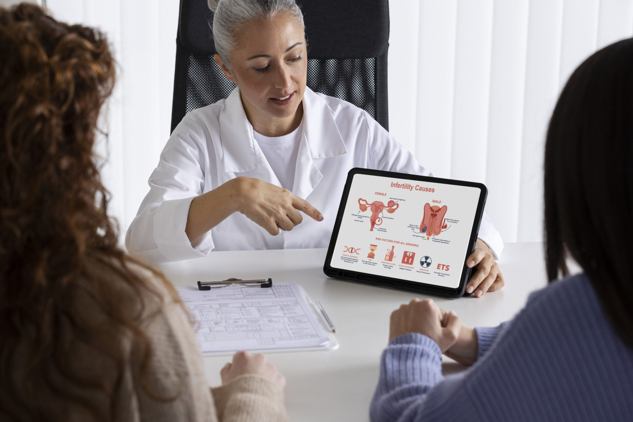
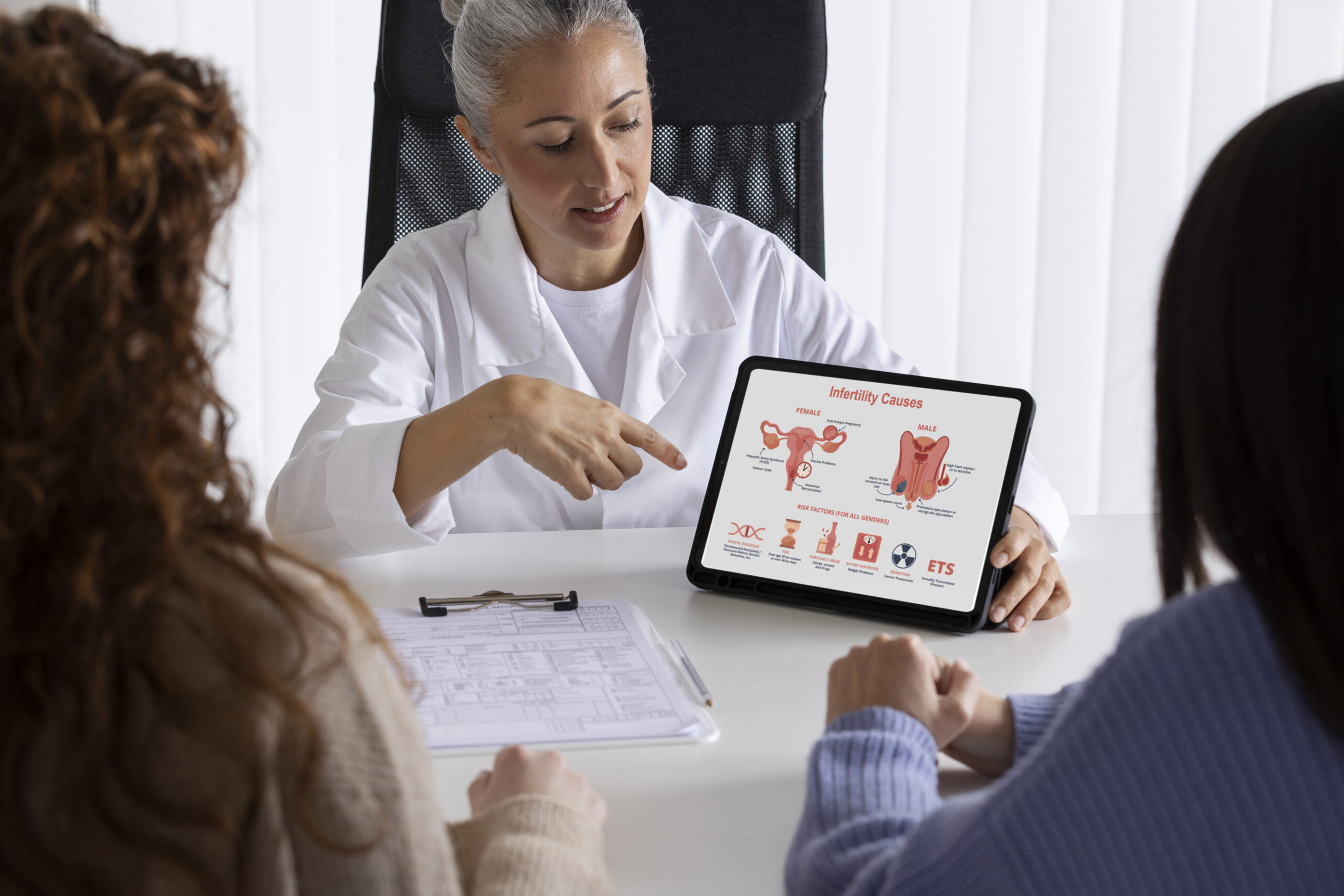
    Infertility is a common medical condition that can also be deeply stressful and emotionally painful for individuals and couples.

    But you are not alone. It is more common than many people realize.

    According to the CDC, 1 in 5 (19%) of married women aged 15 to 49 in the U.S. with no prior births are unable to get pregnant after 1 year of trying.

    Studies suggest that after 1 year of having unprotected sex, 12% to 15% of couples are unable to conceive, and after 2 years, 10% of couples still have not had a live-born baby.

    Infertility is not only a “woman’s problem.”

    Causes may arise from the female side or from the male side.  Many couples have mixed or unexplained factors, which is why evaluating both partners is standard practice.

    Some causes of infertility are preventable. Medications or assisted reproductive technologies are aimed at maximizing the chances of conception.

    In this guide, you’ll gain an understanding of what infertility is, its types, causes, signs and symptoms, diagnoses, and available treatment options.

    What is Infertility?

    what is infertility

    According to the World Health Organization (WHO), infertility is a disease of the male or female reproductive system defined by the failure to achieve a pregnancy after 12 months or more of regular unprotected sexual intercourse.

    Conception requires a series of coordinated steps: a mature egg released during the ovulation portion of the menstrual cycle, sperm reaching and fertilizing the egg (typically in the fallopian tube), formation of an embryo, and successful embryo implantation in a receptive uterine lining.

    Disruption at any step, irregular or absent ovulation (menstrual cycle dysregulation), poor egg quality or reduced ovarian reserve, impaired sperm quality (count, shape, motility), tubal blockage, or uterine factors, can cause infertility.

    What are the Types of Infertility?

    Infertility affects millions of people and has an impact on their families and communities.

    WHO estimates suggest that approximately one in every six people of reproductive age worldwide experiences infertility in their lifetime.

    Types of infertility include:

    1. Primary Infertility

    This refers to a person or couple who have never achieved a pregnancy (or live birth) despite at least 12 months of regular, unprotected intercourse (or 6 months if the woman is 35 or older).

    Because no prior conception has occurred, the evaluation typically begins once the diagnostic threshold (12 or 6 months) is reached.

    Also, there is no prior successful pregnancy history; clinicians often consider a full infertility work-up (ovulation assessment, ovarian reserve, tubal/uterine imaging, semen analysis) more promptly.

    The absence of prior conception may suggest more significant or multiple contributing factors.

    2. Secondary Infertility

    It occurs when a person or couple has had at least one prior pregnancy (regardless of outcome) but is now unable to conceive after the defined interval of regular unprotected intercourse.

    Because a prior conception occurred, some systems (ovulatory, tubal, sperm) were once functional. The evaluation may focus on “what changed.

    For example, age-related decline in ovarian reserve, new tubal damage, and a change in partner sperm quality.

    Therefore, prompt assessment is required, especially if age or other risk factors are present. Still, the perceived urgency is sometimes slightly less than in primary infertility (depending heavily on age and other context).

    3. Female-Factor Infertility

    This refers to cases in which the primary or sole contributing cause lies in the female partner’s reproductive system.

    For example, ovulatory dysfunction (irregular or absent ovulation), diminished ovarian reserve (reduced egg quantity/egg quality), tubal factor (blocked or damaged fallopian tubes), uterine or endometrial factors (uterine fibroids, endometriosis, poor endometrial receptivity).

    The clinician (often a reproductive endocrinologist) will assess menstrual cycle regulation, hormone levels (FSH, AMH), ovulation status, ovarian reserve, tubal patency, or uterine anatomy.

    Because female fertility declines with age (especially egg quality after age 35–40), the timeframe for intervention may be compressed, meaning earlier work-up and possibly earlier referral for assisted reproductive technologies may be indicated when diminished ovarian reserve or significant structural issues are found.

    4. Male-Factor Infertility

    This refers to infertility in which the male partner is the primary contributor. Common issues include reduced sperm quality (count, motility, morphology), sperm transport problems, hormonal/andrologic disorders.

    For this, evaluation begins with a partner’s semen analysis, hormonal testing (testosterone, FSH, LH), a physical/andrologic exam, and possibly imaging or genetic testing.

    Also, management may include lifestyle modifications, medical or surgical treatments (e.g., varicocele repair, hormonal therapy), and assisted reproduction when appropriate.

    If the male factor is severe, the timeline for moving to assisted reproductive technologies may be faster rather than prolonged expectant management.

    5. Tubal Infertility

    This is a specific female-factor category in which the fallopian tubes are blocked, scarred, or damaged, preventing the egg and sperm from meeting or the embryo from reaching the uterus.

    It’s often evaluated via imaging (hysterosalpingogram, sonohysterography, or laparoscopy).

    If tubal damage is significant (e.g., hydrosalpinx, bilateral obstruction), the timeline for moving to assisted reproduction (such as in vitro fertilization) may be accelerated because natural conception is unlikely.

    In some cases, surgical repair may be an option.

    6. Combined Infertility

    This category applies when both partners (female and male) have contributing fertility-limiting factors.

    For example, the female partner may have diminished ovarian reserve or tubal damage, and the male partner may have suboptimal sperm quality.

    Because two sets of problems exist, management often needs coordinated care involving both the reproductive endocrinologist and male fertility specialist (andrologist).

    Also, the timeline for intervention may be shorter given the combined burden, so clinicians often recommend more proactive assessment and treatment rather than “wait and see.”

    7. Unexplained Infertility

    This occurs when, after a standard infertility evaluation (including ovulation assessment, ovarian reserve testing, semen analysis, tubal and uterine imaging), no specific cause is identified.

    Even though no cause is found, treatment is still possible, but it’s empiric.

    Options may include ovulation induction, intrauterine insemination (IUI) timed to the fertile window, or more directly assisted reproductive technologies (ART), depending on age and other risk factors.

    Because there is no targetable cause, prognosis depends heavily on age, duration of infertility, and other subtle factors; timing becomes important, especially when the female partner is older.

    What Causes Infertility?

    what are the causes of infertility

    Many underlying medical, lifestyle, and environmental factors can impair egg quality, sperm quality, or the fertilisation/implantation sequence.

    The reasons for infertility vary from person to person. Here are the most common causes of infertility in women.

    Some reasons for female fertility problems include:

    • Ovulatory Disorders

    A normal menstrual cycle averages about 28 days; cycles that are fairly regular, roughly every 21 to 35 days, usually indicate that ovulation is occurring. Irregular or infrequent periods often mean ovulation is irregular or absent.

    When ovarian function is disrupted, a health-care provider should evaluate possible causes.

    Typical tests include follicle-stimulating hormone (FSH), anti-Müllerian hormone (AMH), and antral follicle count (AFC) on pelvic ultrasound.

    Common conditions that affect ovarian function

    • Polycystic ovary syndrome (PCOS): The most frequent cause of ovulatory infertility. PCOS disrupts the normal hormonal signals that trigger ovulation, causing irregular or absent ovulation and often associated metabolic and androgenic features.
    • Diminished ovarian reserve (DOR): Fewer eggs remain in the ovaries than expected for a woman’s age. DOR may be congenital, related to prior surgery or medical treatment, or unexplained; it reduces both the quantity and often the quality of eggs available for fertilization.
    • Hypothalamic or pituitary dysfunction: Research shows that problems in the brain’s reproductive control centers (the hypothalamus and pituitary) can alter the release of GnRH, LH, and FSH, impairing follicle development and ovulation.
    • Functional hypothalamic amenorrhea (FHA): A reversible condition caused by low body weight, rapid weight loss, excessive exercise, or significant stress. FHA suppresses GnRH secretion and stops or reduces ovulation.
    • Primary ovarian insufficiency (POI): Also called premature ovarian failure, POI occurs when the ovaries stop functioning before age 40. It’s characterized by irregular or absent periods, elevated FSH, and low estrogen, and may result in early loss of fertility.

    If ovarian dysfunction is suspected, timely evaluation and targeted testing help identify the specific cause and guide management, whether lifestyle changes, medical treatment to restore ovulation, or fertility-preserving or assisted-reproductive options.

    • Age-Related Decline

    As a woman ages, two key fertility-related processes deteriorate: the number of eggs remaining in her ovaries (often measured as ovarian reserve) and the ability of those eggs to fertilize and develop into healthy embryos (egg quality).

    Research shows that age-related changes in oocyte (egg) energy metabolism (for example, reduced ATP production, altered mitochondrial and lipid metabolism) degrade egg quality.

    Another study found that the prevalence of Diminished Ovarian Reserve (DOR) increased significantly with age, with more than double the rate in women over 30 compared to those under 30.

    Because both quantity and quality decline with age, the “fertile window” shrinks in both monthly opportunities and cumulative chances.

    • Tubal Issues

    When the tubes are damaged, blocked, scarred, or their epithelial lining is compromised, the egg and sperm cannot meet properly, or the resulting embryo cannot travel safely to the uterus. This condition is known as tubal factor infertility (TFI).

    Causes of fallopian tube damage or blockage can include:

    • Blockage or scarring: Infections trigger inflammation that leads to adhesions, scar tissue, or complete tubal occlusion, preventing the egg and sperm from meeting.
    • Epithelial damage: The ciliated cells of the fallopian tube help transport the fertilised egg into the uterus. Damage to these cells slows or stops embryo transit.
    • Ectopic risk: Because the embryo’s path is compromised, implantation may occur in the tube itself rather than the uterus, leading to ectopic pregnancy.

    One retrospective cohort of 2,892 women with tubal infertility found that those with prior ectopic pregnancy had worse outcomes in IVF/ICSI cycles.

    Because tubal damage is often irreversible, prompt identification can shift management earlier to assisted reproductive technologies (ART) rather than prolonged natural-cycle attempts.

    • Endometriosis

    Endometriosis is the presence of endometrial-like tissue outside the uterus (for example, on the ovaries, fallopian tubes, or pelvic peritoneum).

    This aberrant tissue can cause inflammation, adhesions (scar tissue), hormonal dysregulation, and altered pelvic anatomy. These changes can impact fertility by:

    • Impairing ovulation or reducing egg/follicle quality when the ovary is involved (e.g., endometrioma).
    • Causing tubal/peritoneal adhesions that interfere with the meeting of egg and sperm or embryo transport.
    • Altering the uterine lining (endometrium) so that embryo implantation is less likely.
    • Creating a hostile, inflammatory environment in the pelvis may reduce fertility potential.

    A study found that women with endometriosis exhibit endometrial changes (hormonal imbalances, immune dysregulation, altered microRNA/gene expression) that reduce the likelihood of successful implantation.

    Another research report found that, among women with endometriosis, oxidative stress, dysregulated steroidogenesis (via aromatase P450), and elevated cytokines (IL-6, IL-8, TNF-α) were identified as factors likely impairing follicle/oocyte development and thus fertility.

    • Uterine Factors

    Uterine-factor infertility refers to conditions in the uterus that interfere with one or more steps of conception, implantation, or maintenance of a pregnancy.

    These include anatomical abnormalities and acquired lesions that alter the uterine cavity, lining, or musculature.

    A cross-sectional study found a high prevalence of acquired uterine anomalies (polyps, submucosal fibroids) and congenital anomalies (septate, bicornuate uteri) among sub-fertile women.

    Because these uterine issues affect the “plumbing and lining” part of the reproductive system, the cavity where the embryo must implant, timely detection and repair can often improve chances of conception.

    • Thyroid Disease

    According to the American Thyroid Association, one in eight women will develop a thyroid disorder in her lifetime.

    Thyroid problems usually fall into two main types:

    • Hyperthyroidism (overactive thyroid): The gland makes too much thyroid hormone.
    • Hypothyroidism (underactive thyroid): The gland does not produce enough thyroid hormone.

    Some people may also develop thyroid nodules, which are small lumps or growths in the thyroid. Most nodules are harmless, but in some cases, they can cause hormonal changes or make swallowing uncomfortable.

    Research shows that overt (clinical) hypothyroidism is consistently associated with menstrual irregularities, anovulation, luteal-phase defects, and reduced conception rates. Treating overt hypothyroidism (levothyroxine) typically restores regular cycles and improves chances of conceiving.

    Another research from the National Institutes of Health (NIH) also highlights that about 2–4% of women of reproductive age have hypothyroidism.

    This condition can interfere with normal ovulation and disrupt menstrual cycles, which may lead to infertility. Hypothyroidism can appear on its own, develop after pregnancy, or occur following treatment for an overactive thyroid. In some cases, it may also be present from birth.

    • Autoimmune Disorders

    Autoimmune diseases develop when the immune system mistakenly targets the body’s own healthy cells, leading to ongoing inflammation and tissue damage. This heightened immune response can impact fertility in several ways.

    A large population-based study analyzed over 568,000 births and discovered that women with untreated infertility were 25% more likely to develop conditions such as lupus, Sjögren’s syndrome, or inflammatory myopathies within nine years after delivery.

    Also, the research indicates that infertility itself may act as an early warning sign for underlying autoimmune dysfunction, even when a pregnancy is achieved naturally.

    Autoimmune disorders can affect fertility in several ways:

    • Hormonal disruptions: Some autoimmune conditions can damage hormone-producing glands, leading to irregular periods and ovulation issues.
    • Chronic inflammation: Ongoing inflammation may affect egg quality, embryo development, and the uterus’s ability to accept an embryo.
    • Auto-antibodies: In conditions like antiphospholipid syndrome, antibodies can interfere with blood flow to the uterus and placenta, increasing the risk of miscarriage.
    • Medication effects: Certain immunosuppressants used to treat autoimmune diseases may temporarily reduce fertility or affect ovarian function.
    • Higher miscarriage risk: Women with autoimmune disorders, especially lupus and antiphospholipid syndrome, have a greater chance of pregnancy loss.

    Overall, autoimmune activity, inflammation, and treatment effects can all contribute to fertility challenges.

    • Chronic Medical Conditions

    Long-term illnesses such as diabetes, hypertension, autoimmune disorders, kidney disease, and untreated thyroid problems can disrupt the delicate hormonal balance needed for ovulation and healthy menstrual cycles.

    These conditions may also affect blood flow to reproductive organs, increase inflammation, or lead to complications like irregular periods, poor egg quality, or difficulty maintaining a pregnancy.

    For example, research shows that chronic metabolic disorders such as obesity and metabolic syndrome impair fertility by disrupting the hypothalamic–pituitary–ovarian (HPO) axis, lowering oocyte quality, and reducing implantation and live-birth rates; multiple recent reviews document these mechanisms and worse assisted reproduction outcomes in obese women.

    Another research indicates that decreased ovarian reserve, altered ovarian function, and worse clinical pregnancy rates in women with diabetes.

    • Lifestyle Factors

    Unhealthy habits such as smoking, excessive alcohol use, poor diet, and lack of exercise can disrupt the hormonal balance required for ovulation and regular menstrual cycles.

    Moreover, exposure to environmental toxins, like pesticides, plastics (BPA), and certain chemicals, may also damage eggs and sperm or affect implantation.

    Plus, poor sleep, chronic dehydration, and overuse of caffeine further strain the body’s hormonal regulation.

    What are the Signs of Infertility?

    what are the signs of infertility

    Infertility in people who menstruate is often first suspected from objective clues in the reproductive history, physical exam, or simple tests.

    Many of these signs point to hormonal or structural problems that can impair ovulation, egg quality, tubal function, or uterine receptivity.

    The common signs of infertility include:

    • Irregular menstrual cycles: Cycles shorter than 21 days, longer than 35 days, or unpredictable, often a sign of ovulatory problems like PCOS or hormonal imbalance.
    • Very heavy or very light periods: Abnormal flow can signal fibroids, polyps, hormonal disorders, or uterine lining problems that affect implantation.
    • Absence of ovulation: No ovulation confirmed by tests (like low mid-luteal progesterone). Seen in PCOS, thyroid issues, hyperprolactinemia, or POI.
    • History of pelvic inflammatory disease (PID) or STIs: Documented infections such as chlamydia or gonorrhea increase the risk of tubal scarring and blocked fallopian tubes.
    • Recurrent miscarriages (2 or more): Repeated pregnancy loss may indicate uterine abnormalities, genetic issues, autoimmune disorders, or hormonal imbalance.
    • Abnormal vaginal discharge (infection-related): Clinically confirmed infections can travel upward and damage the tubes, affecting fertility.
    • Persistent pelvic pain: Exam-related or recorded chronic pelvic pain may be a sign of endometriosis or pelvic adhesions that interfere with conception.

    What are the Symptoms of Infertility

    what are the symptoms of infertility

    While infertility itself often has no symptoms, many women experience physical changes that point to an underlying condition affecting their reproductive health.

    Common symptoms linked to infertility include:

    • Painful periods (dysmenorrhea): Severe cramping may indicate endometriosis or pelvic inflammation.
    • Pain during intercourse (dyspareunia): Often linked to endometriosis, pelvic adhesions, or chronic inflammation.
    • Chronic pelvic pain: Persistent pain outside menstruation may reflect endometriosis, fibroids, or pelvic scarring.
    • Hot flashes or night sweats: A sign of low estrogen and possible premature ovarian insufficiency (POI).
    • Nipple discharge (galactorrhea): Can indicate hyperprolactinemia, which disrupts ovulation.
    • Rapid weight gain: Frequently associated with insulin resistance or hormonal imbalance, especially in PCOS.
    • Acne or excessive facial hair (hirsutism): Signs of androgen excess are often seen in PCOS and other ovulatory disorders.

    How Is Infertility Diagnosed?

    No single test can detect every cause of infertility, and many tests miss mild problems.

    A comprehensive approach typically includes:

    Detailed Medical History and Physical Exam

    Your clinician reviews your menstrual cycle, past pregnancies, infections, surgeries, medications, lifestyle factors, and overall health.

    A pelvic exam checks for fibroids, ovarian masses, or signs of endometriosis. This guides which tests come next.

    Basic Ovulation Testing (Is ovulation occurring?)

    Home ovulation kits detect the LH surge and help time intercourse.

    Also, a mid-luteal progesterone test is performed ~7 days before your period to confirm ovulation. Low levels suggest absent or irregular ovulation.

    Ovarian-Reserve Testing (How many eggs remain/ovarian activity)

    It typically includes three key measures:

    • AMH (anti-Müllerian hormone): A blood test that can be done any time in the cycle and provides an estimate of ovarian reserve.
    • FSH and estradiol: Usually measured on cycle day 2–4; an elevated FSH or abnormal estradiol can indicate reduced reserve.
    • Antral follicle count (AFC): A transvaginal ultrasound performed early in the cycle counts small follicles and gives a visual estimate of ovarian reserve.

    Transvaginal ultrasound (TVUS) (structural and functional imaging)

    A transvaginal ultrasound evaluates the uterus and ovaries: it detects fibroids, ovarian cysts or endometriomas, and helps assess the antral follicle count.

    It’s a routine, non-invasive test used throughout the diagnostic workup.

    Tests of Tubal Patency

    To check whether eggs and sperm meet, the following tests are done:

    • Hysterosalpingography (HSG): X-ray with contrast to see if the fallopian tubes are open.
    • Saline infusion sonohysterogram (SIS): ultrasound with saline to detect polyps, fibroids, or adhesions in the uterine cavity.

    Diagnostic Laparoscopy (Selected cases)

    A laparoscopy is a minimally invasive surgery (small incisions, camera into the pelvis) reserved for specific indications: suspected endometriosis not visible on imaging, persistent pelvic pain, or unclear tubal disease after HSG/other tests.

    It can both diagnose and, in some cases, treat adhesions or endometriosis.

    Remember, the testing protocols vary between providers, so it is reasonable to seek a second opinion if you’re unsure about results or next steps.

    Infertility Treatment Options: From Least Invasive to Most Advanced

    infertility treatment options

    Infertility treatments vary in complexity and invasiveness, ranging from lifestyle changes to advanced assisted reproductive technologies (ART). Also, they are tailored to age, diagnosis, and reproductive goals.

    Here are some of the most commonly used infertility treatment options:

    Lifestyle Changes

    Optimizing overall health can naturally improve fertility. This includes achieving a healthy weight, eating a balanced diet, quitting smoking, limiting alcohol, managing stress, and avoiding endocrine-disrupting chemicals.

    Even modest lifestyle adjustments can improve ovulation, egg quality, sperm quality, and endometrial receptivity.

    Ovulation Induction Medications

    For women with ovulatory dysfunction (such as PCOS or hormonal imbalance), medications stimulate ovulation:

    • Clomiphene citrate: Oral medication that induces ovulation.
    • Letrozole: An aromatase inhibitor often preferred for women with PCOS.
    • Gonadotropins: Injectable hormones for more controlled ovarian stimulation.

    Hormonal regulation may also include correcting thyroid, prolactin, or luteal-phase deficiencies.

    Intrauterine Insemination (IUI)

    IUI involves placing washed sperm directly into the uterus around the conception window to increase the chances of fertilization.

    Often combined with ovulation induction. Suitable for mild male factor infertility, unexplained infertility, or cervical issues.

    In Vitro Fertilization (IVF)

    IVF is an ART procedure where eggs are retrieved from the ovaries, fertilized with sperm in a lab, and the resulting embryos are cultured to the blastocyst stage for transfer.

    Embryo grading helps select the best embryos for transfer, maximizing success rates. IVF is used for tubal ligation factor, severe male infertility, or when IUI has failed.

    Intracytoplasmic Sperm Injection (ICSI)

    A specialized form of IVF where a single sperm is injected directly into an egg. Often used for severe male infertility, low sperm count, poor motility, or previous fertilization failure in IVF.

    Donor Gametes

    When a woman’s eggs are of poor quality, or a man has no viable sperm, donor eggs or sperm can be used. This allows fertilization with donor genetic material.

    This is often recommended for women over 40 or for couples where male infertility is severe. Donor eggs have a higher success rate in older women.

    Gestational Surrogacy

    Gestational surrogacy involves another woman carrying a pregnancy for a couple who cannot conceive or carry a pregnancy themselves. The embryo is created through IVF using the eggs and sperm from the intended parents or donors.

    It is typically used when a woman cannot carry a pregnancy due to health conditions or failed IVF cycles.

    Fertility Preservation (Egg/Embryo Freezing)

    Fertility preservation includes freezing eggs or embryos for future use. This is often done by women who wish to delay childbearing for personal or medical reasons, such as cancer treatment.

    Common for women over 35 who want to delay childbearing, or for those facing medical treatments that may affect fertility.

    However, for men, sperm freezing is an option before undergoing treatments like chemotherapy.

    Choosing the right treatment requires a personalized evaluation by a reproductive endocrinologist or fertility specialist, taking into account age, diagnosis, and prior response to therapy.

    Complications of Infertility Treatment

    Infertility treatments, especially those involving hormonal stimulation or assisted reproduction, carry some risks, though serious problems are relatively rare.

    Here are some complications of infertility treatment:

    Ovarian Hyperstimulation Syndrome (OHSS)

    OHSS occurs when the ovaries over-respond to stimulation, causing fluid leakage, ovarian swelling, and sometimes more severe effects.

    A recent large review estimated that moderate to severe OHSS affects 1–5% of IVF cycles. Severe OHSS can lead to complications such as hemoconcentration, clotting risk, and more.

    Multiple Pregnancy Risk

    Fertility treatments (especially when more than one embryo is transferred, or with strong ovulation stimulation) increase the likelihood of twins or higher-order multiples.

    Also, multiple gestations are a major contributor to pregnancy complications after fertility treatment. Studies show that many of the excess risks (preterm birth, cesarean section, preeclampsia) are mediated by having multiples.

    Ectopic Pregnancy

    Although relatively uncommon, ectopic pregnancy (when an embryo implants outside the uterus) can occur following fertility treatment.

    Research shows that ectopic pregnancies have been observed as one of the more serious adverse events, especially in protocols involving embryo transfer.

    Emotional Stress, Anxiety, Depression

    The emotional toll of infertility treatments can be significant, leading to increased stress, anxiety, and depression.

    The uncertainty of treatment outcomes, coupled with hormonal changes, can exacerbate these feelings.

    For this, support from counselors, support groups, and a strong partner relationship can help alleviate emotional distress.

    Procedure-Related Discomfort

    Some infertility treatments, particularly egg retrieval and sperm collection procedures, may cause temporary discomfort or pain.

    These procedures are generally quick and performed under sedation or local anesthesia, ensuring patient comfort.

    Financial and Insurance Considerations

    Infertility treatments can be expensive, and not all insurance plans cover the full cost.

    This financial burden can add stress to the process, but many fertility clinics offer payment plans, and some regions have specific fertility treatment insurance coverage.

    Medication Side Effects

    Fertility drugs (gonadotropins, hCG trigger, etc.) can cause side effects like bloating, nausea, mood changes, and fluid shifts.

    Because some protocols involve high doses of hormones, the body’s response can be quite variable, which is why close monitoring is essential.

    While there are potential complications associated with infertility treatments, most are rare and manageable with appropriate medical supervision.

    To address these, open communication with healthcare providers and proper monitoring significantly reduce the chances of severe complications.

    How to Prevent Infertility?

    While not all cases of infertility are preventable, many risk factors are modifiable, and taking proactive steps to address these factors can improve your chances of maintaining fertility.

    Here are some practical ways to reduce your risk:

    • Maintain a Healthy BMI: A healthy weight supports regular menstrual cycles and ovulation.
    • Quit Smoking: Smoking affects egg quality in women and sperm in men, reducing fertility.
    • Limit Alcohol Intake: Excess alcohol can disrupt hormone production and ovulation.
    • Use STI Protection: Condoms and regular screenings can prevent infections that damage reproductive organs.
    • Treat Infections Promptly: Prompt treatment of infections such as pelvic inflammatory disease (PID) prevents fertility issues.
    • Manage Thyroid Disease: Regular monitoring and treatment of thyroid issues support fertility.
    • Control Chronic Conditions: Managing diabetes and autoimmune disorders reduces their impact on fertility.
    • Reduce Toxin Exposure: Limiting exposure to BPA and pesticides protects reproductive health.
    • Get Regular Well-Woman Exams: Regular exams help detect fertility issues early.
    • Track Menstrual Cycles: Monitoring your cycle helps identify irregularities that may affect fertility.

    By taking these steps, individuals can improve their reproductive health and increase their chances of conception.

    Frequently Asked Questions (FAQs)

    Why am I not getting pregnant?

    Not getting pregnant can be frustrating, but it is often related to a combination of factors involving both partners. Several factors can affect fertility:

    • Ovulation Issues: Irregular ovulation can prevent conception. Tracking ovulation can help identify this issue.
    • Male-Factor Infertility: Low sperm count or poor motility can impact fertility. A semen analysis can identify sperm issues.
    • Age: As you age, egg quality and quantity decline, reducing the chances of pregnancy.
    • Timing: Conception is most likely when intercourse happens close to ovulation.
    • Underlying Conditions: Conditions like thyroid disorders or diabetes can affect fertility.

    How to know if you’re infertile?

    You may consider a fertility evaluation if you haven’t conceived after 12 months of trying (<35 years) or 6 months (≥35 years). Earlier assessment is recommended for signs like irregular periods, missed ovulation (anovulation), recurrent miscarriages, or known reproductive conditions. Tests often include cycle monitoring, ovulation tracking, and AMH testing to assess ovarian reserve and guide next steps.

    How common is infertility?

    Infertility affects a significant number of couples in the U.S. According to the CDC, about 19% of married women aged 15–49 with no prior births are unable to get pregnant after one year of trying. Delays in childbearing (women starting families later) and demographic shifts contribute to this trend.

    Is infertility genetic?

    Yes, genetics can play a role in certain infertility conditions. Some women may inherit risks for endometriosis, premature ovarian insufficiency (POI), or polycystic ovary syndrome (PCOS), while men can inherit Y-chromosome deletions or Klinefelter syndrome, affecting sperm production. Therefore, a family history of reproductive issues can signal a higher risk. Couples concerned about hereditary infertility may benefit from genetic counseling to understand risks and testing options.

    Can infertility be cured?

    Some infertility causes are treatable; for example, hormonal imbalances can be managed with medications, ovulation issues may improve with lifestyle changes, and structural problems like fibroids or tubal blockages can sometimes be corrected surgically. Other cases may not be fully “curable” but can be managed effectively with assisted reproductive technologies (ART) such as IVF. Success depends on the underlying cause, age, and overall reproductive health.

    Conclusion

    Infertility is a common medical condition, affecting nearly 1 in 5 women of reproductive age in the U.S., and often involves factors from both partners.

    Causes range from ovulation disorders, tubal or uterine issues, and age-related egg decline to male-factor infertility, including low sperm count or genetic conditions.

    Many signs, such as irregular cycles, recurrent miscarriages, or difficulty conceiving despite regular intercourse, can signal the need for evaluation. Plus, symptoms like painful periods or hormonal changes may indicate underlying disorders.

    The good news is that most infertility causes are treatable or manageable. Lifestyle changes, ovulation induction, hormonal therapy, surgical correction, or advanced ART procedures like IVF and ICSI can significantly improve the chance of conception.

    While the journey can be emotionally challenging, understanding the causes, monitoring cycles, and accessing appropriate care empowers couples.

    Infertility is not a reflection of personal failure, and with timely support and evidence-based treatment, many individuals and couples achieve successful pregnancy outcomes. Your fertility matters, and help is available.

  • Comparing Fibroid Treatments: Why Minimally Invasive Options Are Changing the Game

    Comparing Fibroid Treatments: Why Minimally Invasive Options Are Changing the Game

    Not long ago, women with problematic fibroids faced a limited and often daunting set of treatment options: suffer through symptoms, undergo major surgery, or accept hysterectomy as the “final solution.” Today’s landscape looks dramatically different, with minimally invasive procedures offering effective relief without the risks and recovery time of traditional surgery.

    At Seamless Medical Centers, we’ve witnessed this evolution firsthand. Our patients often express amazement that they waited so long to seek treatment, not realizing that cutting edge solutions could address their symptoms with minimal disruption to their lives.

    The Traditional Treatment Landscape

    Historically, fibroid treatment followed a predictable progression:

    Watchful Waiting: Monitoring fibroids without active treatment

    Medical Management: Hormonal therapies to control symptoms

    Myomectomy: Surgical removal of fibroids while preserving the uterusHysterectomy: Complete removal of the uterus

    While these approaches remain valid for specific situations, they often involve significant limitations. According to the Fibroid Foundation, 300,000 women a year have hysterectomies due to uterine fibroids, representing a major surgical solution for what might be addressed less invasively.

    The Minimally Invasive Revolution

    Modern interventional radiology has transformed uterine fibroid treatment by offering procedures that are:

    • Less Invasive: Using small incisions or no incisions at all
    • More Precise: Targeting fibroids while preserving healthy tissue
    • Faster Recovery: Returning to normal activities within days, not weeks
    • Outpatient-Based: Going home the same day rather than hospital stays

    Detailed Treatment Comparison

    Traditional Open Myomectomy

    Approach: Large abdominal incision to remove fibroids

    Recovery: 4-6 weeks off work

    Hospital Stay: 2-3 days typically

    Risks: Higher infection risk, significant scarring, longer anesthesia exposure

    Laparoscopic/Robotic Myomectomy

    Approach: Multiple small incisions with specialized instruments

    Recovery: 2-3 weeks off work

    Hospital Stay: Usually same-day or overnight

    Advantages: Smaller scars, reduced pain compared to open surgery

    Hysterectomy

    Approach: Removal of entire uterus (various surgical approaches)

    Recovery: 6-8 weeks for full recovery

    Considerations: Permanent solution but eliminates future pregnancy possibility

    Impact: Significant life change requiring careful consideration

    Radiofrequency Ablation (RFA)

    Approach: Targeted thermal energy to destroy fibroid tissue

    Recovery: 3-5 days average return to work

    Hospital Stay: Outpatient procedure

    Precision: Treats fibroids while preserving healthy tissue

    Uterine Fibroid Embolization (UFE) – Our Specialty

    Approach: Blocking blood supply to fibroids through catheter in leg artery

    Recovery: About 1 week

    Performed by: Interventional radiologist

    Considerations: UFE has proven to be safe, versatile, and effective in reducing or eliminating symptoms related to uterine fibroids

    Why Minimally Invasive Matters

    The advantages of minimally invasive approaches extend beyond shorter recovery times:

    Reduced Complications: Smaller incisions mean lower infection risks and fewer surgical complications.

    Faster Healing: Less tissue trauma allows your body to heal more quickly and with less pain.

    Preserved Function: Maintaining uterine structure and function when possible offers long-term benefits.

    Cost Effectiveness: Shorter procedures, same-day discharge, and faster return to work reduce overall healthcare costs.

    Quality of Life: Minimal downtime means less disruption to work, family, and personal activities.

    Real-World Outcomes

    Research consistently demonstrates the effectiveness of modern minimally invasive approaches. Studies show UFE treatments were associated with fibroid volume reduction of 46.0% at 3 months and 65.4% at 12 months, with significant improvements in quality of life scores.

    Patient satisfaction rates for minimally invasive procedures consistently exceed those for traditional surgical approaches, largely due to the combination of effective symptom relief and reduced treatment burden.

    Choosing the Right Approach

    The best treatment depends on multiple factors:

    Fibroid Characteristics: Size, location, and number of fibroids influence treatment options

    Symptom Profile: Heavy bleeding, pressure symptoms, or pain may favor different approaches

    Personal Goals: Desire to preserve fertility, avoid surgery, or minimize recovery time

    Medical History: Previous surgeries, overall health, and other medical conditions

    Lifestyle Factors: Work demands, family responsibilities, and personal preferences

    Seamless Medical Centers Advantage

    Our focus on minimally invasive interventional radiology offers several key benefits:

    Expertise: Board-certified interventional radiologists with extensive experience in image-guided procedures

    Technology: State-of-the-art equipment and advanced imaging for optimal precision and safety

    Comprehensive Care: From initial consultation through follow-up, we support you throughout your treatment journey

    Insurance Coverage: Most major insurance plans cover our procedures, and we handle authorization processes

    Making an Informed Decision

    Choosing fibroid treatment is a personal decision that should be based on accurate, current information about all available options. Many women are surprised to learn about minimally invasive alternatives to traditional surgery.

    During your consultation, we’ll:

    • Review your specific fibroid characteristics
    • Discuss your symptoms and treatment goals
    • Explain all appropriate treatment options
    • Provide detailed information about what to expect
    • Answer all your questions thoroughly

    The Future Is Now

    The landscape of fibroid treatment continues to evolve, with new technologies and techniques constantly emerging. However, the fundamental shift toward minimally invasive, precision-based treatments represents a permanent change in how we approach this common condition.

    Ready to explore your options?

    Contact Seamless Medical Centers to learn how modern, minimally invasive treatments can address your fibroid symptoms effectively while minimizing disruption to your life. Your journey to better health starts with understanding your choices.